

Diagnosis and management of the neurological complications of falciparum malaria
- PMID: 19347024
- PMCID: PMC2859240
- DOI: 10.1038/nrneurol.2009.23
Diagnosis and management of the neurological complications of falciparum malaria
Abstract
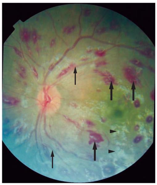
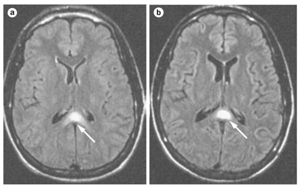
Malaria is a major public health problem in the developing world owing to its high rates of morbidity and mortality. Of all the malarial parasites that infect humans, Plasmodium falciparum is most commonly associated with neurological complications, which manifest as agitation, psychosis, seizures, impaired consciousness and coma (cerebral malaria). Cerebral malaria is the most severe neurological complication; the condition is associated with mortality of 15-20%, and a substantial proportion of individuals with this condition develop neurocognitive sequelae. In this Review, we describe the various neurological complications encountered in malaria, discuss the underlying pathogenesis, and outline current management strategies for these complications. Furthermore, we discuss the role of adjunctive therapies in improving outcome.
Figures
References
-
- Mishra SK, Mohanty S, Satpathy SK, Mohapatra DN. Cerebral malaria in adults—a description of 526 cases admitted to Ispat General Hospital in Rourkela, India. Ann. Trop. Med. Parasitol. 2007;101:187–193. - PubMed
-
- Newton CR, Warrell DA. Neurological manifestations of falciparum malaria. Ann. Neurol. 1998;43:695–702. - PubMed
-
- Severe falciparum malaria. World Health Organization, Communicable Diseases Cluster. Trans. R. Soc. Trop. Med. Hyg. 2000;94(Suppl. 1):S1–S90. [No authors listed] - PubMed
Publication types
MeSH terms
Grants and funding
LinkOut - more resources
Full Text Sources
Other Literature Sources
Medical
