. 2009;4(4):e5134.
doi: 10.1371/journal.pone.0005134. Epub 2009 Apr 6.

Molecular phenotypes distinguish patients with relatively stable from progressive idiopathic pulmonary fibrosis (IPF)

Affiliations

Molecular phenotypes distinguish patients with relatively stable from progressive idiopathic pulmonary fibrosis (IPF)

Kathy Boon et al. PLoS One. 2009.

Abstract

Background: Idiopathic pulmonary fibrosis (IPF) is a progressive, chronic interstitial lung disease that is unresponsive to current therapy and often leads to death. However, the rate of disease progression differs among patients. We hypothesized that comparing the gene expression profiles between patients with stable disease and those in which the disease progressed rapidly will lead to biomarker discovery and contribute to the understanding of disease pathogenesis.

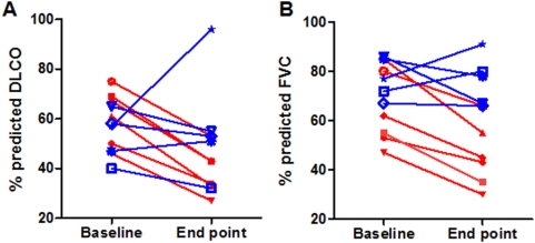
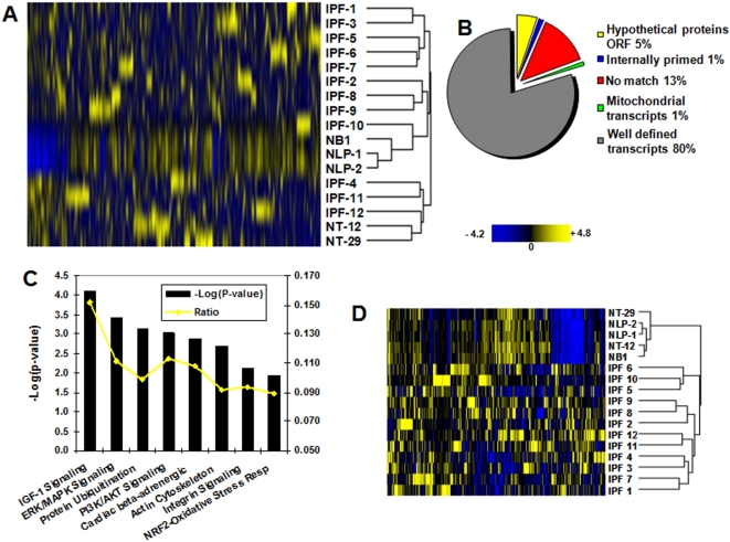
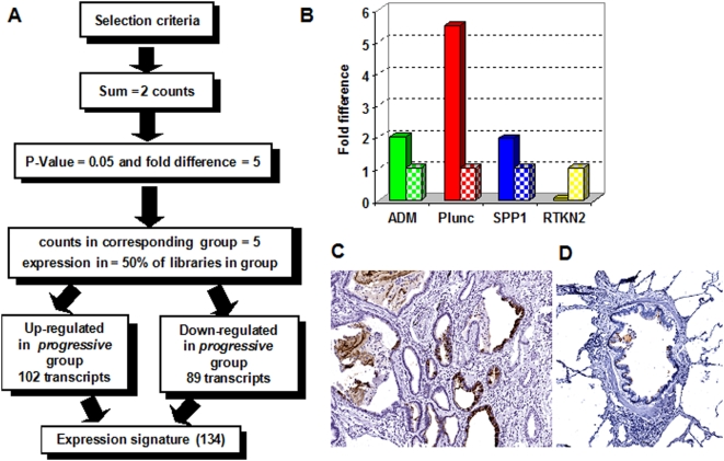
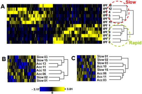
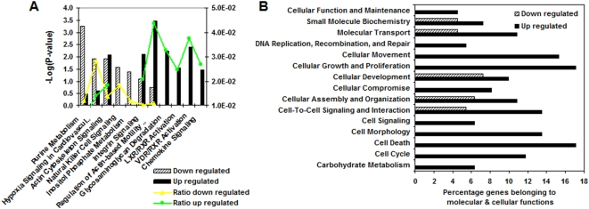
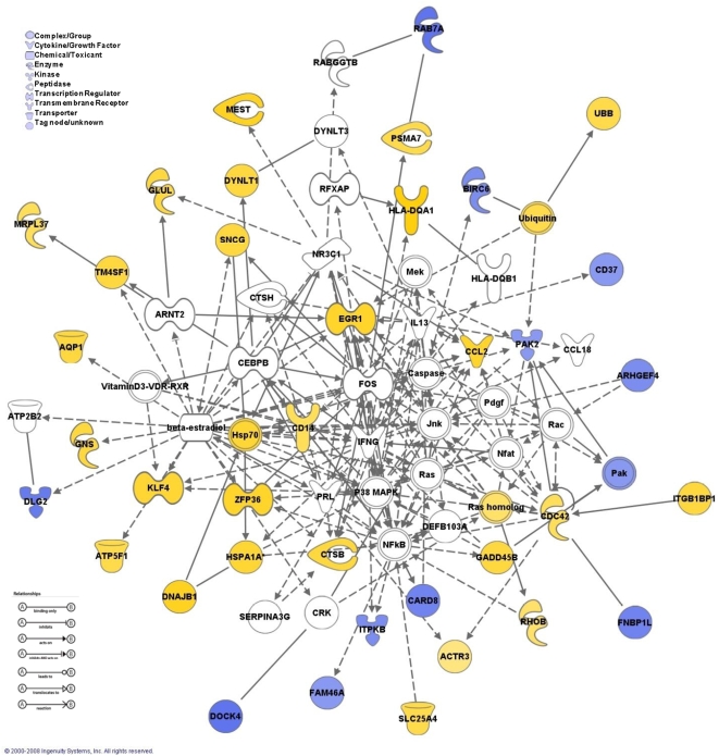
Methodology and principal findings: To begin to address this hypothesis, we applied Serial Analysis of Gene Expression (SAGE) to generate lung expression profiles from diagnostic surgical lung biopsies in 6 individuals with relatively stable (or slowly progressive) IPF and 6 individuals with progressive IPF (based on changes in DLCO and FVC over 12 months). Our results indicate that this comprehensive lung IPF SAGE transcriptome is distinct from normal lung tissue and other chronic lung diseases. To identify candidate markers of disease progression, we compared the IPF SAGE profiles in stable and progressive disease, and identified a set of 102 transcripts that were at least 5-fold up regulated and a set of 89 transcripts that were at least 5-fold down regulated in the progressive group (P-value</=0.05). The over expressed genes included surfactant protein A1, two members of the MAPK-EGR-1-HSP70 pathway that regulate cigarette-smoke induced inflammation, and Plunc (palate, lung and nasal epithelium associated), a gene not previously implicated in IPF. Interestingly, 26 of the up regulated genes are also increased in lung adenocarcinomas and have low or no expression in normal lung tissue. More importantly, we defined a SAGE molecular expression signature of 134 transcripts that sufficiently distinguished relatively stable from progressive IPF.

Conclusions: These findings indicate that molecular signatures from lung parenchyma at the time of diagnosis could prove helpful in predicting the likelihood of disease progression or possibly understanding the biological activity of IPF.


Conflict of interest statement

Competing Interests: The authors have declared that no competing interests exist.

Figures

Figure 1
Figure 1. Forced vital capacity (FVC) and carbon monoxide diffusing capacity (DLCO) values.
The percent predicted DLCO (A) and FVC (B) values are indicated at baseline and end point for the two IPF disease groups. The progressive group is represented in red and the relatively stable group in blue.
Figure 2
Figure 2. Analysis of the IPF transcriptome.
(A) Hierarchical clustering based on the gene expression profiles of 12 IPF and 5 normal lung SAGE libraries described in Table S1. The branch length in the dendrogram represents the distance or relatedness between the samples; the shorter the branch the higher the similarity between samples. In yellow are indicated up regulated and in blue down regulated genes. (B) Tag to gene mapping classification of the 1,121 transcript tags significantly over expressed in IPF when compared to normal lung parenchyma. (C) Most significant canonical pathways associated with pulmonary fibrosis according to the IPA pathway analysis tool. The significance of the association between the dataset and the canonical pathway was measured as a ratio (number of genes from the dataset that map to the pathway divided by the total number of molecules that exist in the canonical pathway). A Fischer's exact test was used to calculate a P-value. (D) Hierarchical clustering based on the 293 transcriptional signature that distinguished IPF from normal lung parenchyma.
Figure 3
Figure 3. Differentially expressed genes in the lung parenchyma from the relatively stable and progressive IPF.
(A) Selection criteria applied in order to find significantly differentially expressed genes. (B) Relative mRNA expression of selected genes. Real-time PCR reactions were performed in triplicate, and the threshold cycle numbers were averaged. Gene expression levels were normalized to GAPDH, and PGK1. The genes ADM (adrenomedullin), Plunc (palate, lung and nasal epithelium carcinoma associated), and SPP1 (osteopontin) were selected as up regulated; and RTKN2 (rhotekin 2) as down regulated in the progressive group. The values obtained for the relatively stable group was arbitrarily set to one to calculate a fold difference. The fold difference in the progressive group is indicated by solid bars and the levels in the relatively stable group are represented by the patterned bars. The differences were not significant as calculated by a Mann-Whitney test. (C) Paraffin-embedded tissue was stained with Plunc antibodies and counterstained with hematoxylin. A representative IPF sample shows strong staining of the secretory/goblet type of bronchial columnar cells (10X magnification). (D) Control bronchial normal lung tissue showed no staining (10X magnification).
Figure 4
Figure 4. Heat map SAGE molecular signature.
(A) Unsupervised clustering of gene expression patterns of IPF lung SAGE libraries described in Table S1, based on the expression signature of 134 transcripts showing a clear distinction between relatively stable (slow) and progressive (rapid) IPF. (B–C) Hierarchical clustering of 8 IPF samples previously identified as a slow and accelerated variants based on 90 (B) or 58 (C) genes in common with the SAGE 134 molecular signature.
Figure 5
Figure 5. Biological differences between progressive and relatively stable disease groups in IPF.
(A) Ingenuity Canonical Pathway analysis showing the most significant pathways associated with the datasets of up and down regulated genes in the progressive group. The significance of the association between the dataset and the canonical pathway was measured as a ratio (number of genes from the dataset that map to the pathway divided by the total number of molecules that exist in the canonical pathway). A Fischer's exact test was used to calculate a P-value. (B) Main molecular and cellular functions significantly associated with the datasets of up and down regulated genes in the progressive group according to the IPA functional analysis tool.
Figure 6
Figure 6. Network analysis.
The network map represents the interaction between members of two networks highlighting the crosstalk between the multiple differentially expressed genes in the progressive group. Nodes represent genes, and theirs shapes represent the functional classes of the gene products. Solid lines indicate a direct interaction and dashed lines indicate an indirect interaction.

References

    1. ATS Dyspnea. Mechanisms, Assessment, and Management: A Consensus Statement. Am J Respir Crit Care Med. 1999;159:321–340. - PubMed
    1. ATS American Thoracic Society/European Respiratory Society International Multidisciplinary Consensus Classification of the Idiopathic Interstitial Pneumonias. This Joint Statement of the American Thoracic Society (ATS), and the European Respiratory Society (ERS) was adopted by the ATS Board of Directors, June 2001 and by The ERS Executive Committee, June 2001. Am J Respir Crit Care Med. 2002;165:277–304. - PubMed
    1. Steele MP, Speer MC, Loyd JE, Brown KK, Herron A, et al. Clinical and Pathologic Features of Familial Interstitial Pneumonia. Am J Respir Crit Care Med. 2005;172:1146–1152. - PMC - PubMed
    1. Gharaee-Kermani M, Gyetko M, Hu B, Phan S. New Insights into the Pathogenesis and Treatment of Idiopathic Pulmonary Fibrosis: A Potential Role for Stem Cells in the Lung Parenchyma and Implications for Therapy. Pharmaceutical Research. 2007;24:819–841. - PubMed
    1. Maher TM, Wells AU, Laurent GJ. Idiopathic pulmonary fibrosis: multiple causes and multiple mechanisms? Eur Respir J. 2007;30:835–839. - PubMed