. 2009 Jun 2;48(21):4548-56.
doi: 10.1021/bi9002887.

Role of loop-loop interactions in coordinating motions and enzymatic function in triosephosphate isomerase

Affiliations

Role of loop-loop interactions in coordinating motions and enzymatic function in triosephosphate isomerase

Yan Wang et al. Biochemistry. .

Abstract

The enzyme triosephosphate isomerase (TIM) has been used as a model system for understanding the relationship between protein sequence, structure, and biological function. The sequence of the active site loop (loop 6) in TIM is directly correlated with a conserved motif in loop 7. Replacement of loop 7 of chicken TIM with the corresponding loop 7 sequence from an archaeal homologue caused a 10(2)-fold loss in enzymatic activity, a decrease in substrate binding affinity, and a decrease in thermal stability. Isotope exchange studies performed by one-dimensional (1)H NMR showed that the substrate-derived proton in the enzyme is more susceptible to solvent exchange for DHAP formation in the loop 7 mutant than for WT TIM. TROSY-Hahn Echo and TROSY-selected R(1rho) experiments indicate that upon mutation of loop 7, the chemical exchange rate for active site loop motion is nearly doubled and that the coordinated motion of loop 6 is reduced relative to that of the WT. Temperature dependent NMR experiments show differing activation energies for the N- and C-terminal hinges in this mutant enzyme. Together, these data suggest that interactions between loop 6 and loop 7 are necessary to provide the proper chemical context for the enzymatic reaction to occur and that the interactions play a significant role in modulating the chemical dynamics near the active site.


Figures
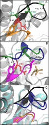
Figure 1
Figure 1
Cartoon rendering of loop structures in cTIM. (A) Open conformation of apo cTIM. Loop 6 is colored black, loop 7 in orange, E165 in red, A169, G173 and A176 in green. Hydrogen bonds between the two loops are shown as black dashed lines, with corresponding distances indicated in black. (B) Closed conformation of bound cTIM using the same color scheme of Figure 1A except for Loop 6, which is colored blue, loop 7 is magenta and phosphoglycolo-hydroxamate (PGH) (27) in brown. (C) Overlay of open (gray) and closed (cyan) conformations of cTIM. Loops are colored in the same scheme of Figure 1A and 1B.
Figure 2
Figure 2
Thermal denaturation of cTIM. CD melting curves of WT (black) and TGAG (red) cTIM. θ is monitored at 214 nm at dimeric protein concentrations of 4 μM. The results shown are the average of three repeat measurements for each enzyme.
Scheme 1
Scheme 1
Reaction of protonated GAP with TIM in 100% D2O. Upon enolization the C2 proton removed by E165 can be directly placed at C1 to form protonated DHAP, exchanged with solvent to form C1 deuterated DHAP (d-DHAP), or exchange with solvent and become C2 deuterated to form deuterated GAP (d-GAP). Substrate derived protons are shown in circles whereas solvent derived deuterons are depicted in squares.
Figure 3
Figure 3
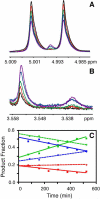
Results of isotope exchange studies. Panels A-B: 1H NMR spectra from the isomerization reaction catalyzed by TGAG mutant TIM. Representative regions of the spectra for GAP hydrate (A), and (B) the DHAP hydrate are shown for the following times after addition of enzyme: 0 min (black), 82 min (red), 156 min (green), 302 min (blue), 522 min (purple). In C: Product distributions of d-GAP (red), d-DHAP (green), and DHAP (blue) as a function of time for wild-type TIM (closed squares, solid lines) and TGAG (open circles, dashed lines).
Figure 4
Figure 4
Chemical shift changes upon mutation of loop 7. (A) Superimposed 1H-15N TROSY HSQC spectra of WT (blue) and TGAG (red) cTIM at 298K and 14.1 T, with panels for close-up views of selected loop 6 residues. (B) Composite chemical shift changes between WT and TGAG cTIM as a function of amino acid sequence. Composite chemical shift changes were calculated using the equation ((ΔδHN2+ΔδN225)2).
Figure 5
Figure 5
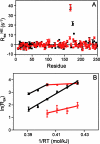
Chemical exchange contribution to R2β. (A) Residue-specific chemical exchange contribution to R2β of WT (black) and TGAG (red) cTIM at 298 K. (B) Temperature dependent relationship of Rex for V167 (■) and T177 (□) in WT (black) and TGAG (red) cTIM. The activation energy for loop closure is extracted from the slope of the linear regression fit to the data points.
Figure 6
Figure 6
Conformational exchange in cTIM. TROSY-selected R1ρ dispersion curves for V167 (top) and T177 (bottom) in WT (black) and TGAG (red) cTIM measured at 298K.

Similar articles

Cited by

References

    1. Hedstrom L, Szilagyi L, Rutter WJ. Converting trypsin to chymotrypsin: the role of surface loops. Science. 1992;255:1249–1253. - PubMed
    1. Peng T, Zintsmaster JS, Namanja AT, Peng JW. Sequence-specific dynamics modulate recognition specificity in WW domains. Nat Struct Mol Biol. 2007;14:325–331. - PubMed
    1. Venkitakrishnan RP, Zaborowski E, McElheny D, Benkovic SJ, Dyson HJ, Wright PE. Conformational changes in the active site loops of dihydrofolate reductase during the catalytic cycle. Biochemistry. 2004;43:16046–16055. - PubMed
    1. Wang L, Pang Y, Holder T, Brender JR, Kurochkin AV, Zuiderweg ER. Functional dynamics in the active site of the ribonuclease binase. Proc. Natl. Acad. Sci. U S A. 2001;98:7684–7689. - PMC - PubMed
    1. Watt ED, Shimada H, Kovrigin EL, Loria JP. The mechanism of rate-limiting motions in enzyme function. Proc Natl Acad Sci U S A. 2007;104:11981–11986. - PMC - PubMed

Publication types