. 2009 Jun 19;4(6):447-56.
doi: 10.1021/cb900021q.

Structure-based design of a periplasmic binding protein antagonist that prevents domain closure

Affiliations

Structure-based design of a periplasmic binding protein antagonist that prevents domain closure

M Jack Borrok et al. ACS Chem Biol. .

Abstract

Many receptors undergo ligand-induced conformational changes to initiate signal transduction. Periplasmic binding proteins (PBPs) are bacterial receptors that exhibit dramatic conformational changes upon ligand binding. These proteins mediate a wide variety of fundamental processes including transport, chemotaxis, and quorum sensing. Despite the importance of these receptors, no PBP antagonists have been identified and characterized. In this study, we identify 3-O-methyl-d-glucose as an antagonist of glucose/galactose-binding protein and demonstrate that it inhibits glucose chemotaxis in E. coli. Using small-angle X-ray scattering and X-ray crystallography, we show that this antagonist acts as a wedge. It prevents the large-scale domain closure that gives rise to the active signaling state. Guided by these results and the structures of open and closed glucose/galactose-binding protein, we designed and synthesized an antagonist composed of two linked glucose residues. These findings provide a blueprint for the design of new bacterial PBP inhibitors. Given the key role of PBPs in microbial physiology, we anticipate that PBP antagonists will have widespread uses as probes and antimicrobial agents.


Figures
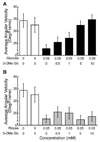
Figure 1
Figure 1
The compound 3-OMe Glc inhibits chemotaxis toward glucose but not ribose. Motion analysis of wild-type E. coli (AW607) upon treatment with glucose (A) or ribose (B) in the presence of increasing concentrations of 3-OMe Glc. Motion analysis was performed on at least 3 independent experiments of 6–8 s duration. Videos were recorded within 45 s of stimulant addition. Error bars are given in 2σ uncertainties.
Figure 2
Figure 2
The complex of 3-OMe Glc and GGBP is in an open conformation. (A) SAXS data (dotted lines) from unliganded GGBP (black), 3-OMe Glc- bound GGBP (cyan), or Glc-bound GGBP (green). Linear fits of these data (solid lines) in the 0.021 > Q > 0.046 region were used to determine slope and Rg. (B) 3-OMe Glc (cyan sticks) binds to the C-terminal domain of the open conformation of GGBP (brown ribbons). Side chains of Tyr10 and Asp14 (shown as brown sticks) would undergo steric clashes upon closure. (C) A superposition of the C-terminal domain of the 3-OMe Glc-bound complex onto the C-terminal domain of the Glc-bound structure. The depiction highlights steric clashes with side chains (cyan sticks with calculated molecular surface) that prevent formation of the closed signaling state of GGBP when 3-OMe Glc binds.
Figure 3
Figure 3
The structure of 3-OMe Glc bound to GGBP determined by x-ray crystallography. (A) An Fo-Fc map (cyan mesh, contoured at 2.5 σ) was generated with 3-OMe Glc (cyan) and HOH 597 omitted. (B) Glucose (green) bound to GGBP (PDB ID: 2FVY) is depicted, along with putative hydrogen bonds to selected residues in the C terminal domain cleft. Complete hydrogen bonding networks for 3-OMe Glc (C) and glucose (D) are illustrated.
Figure 4
Figure 4
Design strategy for an inhibitor that stabilizes the open form of GGBP (A) Structures of closed, glucose-bound GGBP (2FVY) and open, unliganded GGBP (2FW0) were used to generate an unbound model with one glucose molecule occupying each side of the binding cleft. (B) In this model, many stacking and hydrogen bonding interactions in the closed form are maintained (black dashes). (C) Two glucose molecules can be covalently linked at the 1 and 3 positions with an ethylene tether to form the dimeric wedge inhibitor (DWI, 3-O-(2'-beta-D-glucopyanosyloxyethyl)-D-glucose). (D) Chemotactic responses to glucose in E. coli (AW607) cells were inhibited by the DWI. Motion analysis error bars are given in 2σ uncertainties.
Figure 5
Figure 5
Structures of other PBPs indicate that our antagonist design strategy is broadly applicable. Open, unbound and closed, ligand-bound structures of the quorum sensing PBPs LsrB (green, PDB codes 1TJY and 1TM2) and LuxP (yellow, PDB codes 1JX6 and 1ZHH) were used to generate open models with two AI-2 molecules bound to each cleft.

References

    1. Tam R, Saier MH. Structural, functional, and evolutionary relationships among extracellular solute-binding receptors of bacteria. Microbiol. Rev. 1993;57:320–346. - PMC - PubMed
    1. Chen X, Schauder S, Potier N, Van Dorsselaer A, Pelczer I, Bassler BL, Hughson FM. Structural identification of a bacterial quorum-sensing signal containing boron. Nature. 2002;415:545–549. - PubMed
    1. Quiocho FA, Ledvina PS. Atomic structure and specificity of bacterial periplasmic receptors for active transport and chemotaxis: Variation of common themes. Mol. Microbiol. 1996;20:17–25. - PubMed
    1. Surin BP, Rosenberg H, Cox GB. Phosphate specific transport system of Escherichia coli nucleotide sequence and gene polypeptide relationships. J. Bacteriol. 1985;161:189–198. - PMC - PubMed
    1. Felder CB, Graul RC, Lee AY, Merkle HP, Sadee W. The Venus flytrap of periplasmic binding proteins: an ancient protein module present in multiple drug receptors. AAPS PharmSci. 1999;1:E2. - PMC - PubMed

Publication types

MeSH terms