. 2009 Apr 8;96(7):2577-88.
doi: 10.1016/j.bpj.2008.12.3952.

Mechanism and energetics of charybdotoxin unbinding from a potassium channel from molecular dynamics simulations

Affiliations

Mechanism and energetics of charybdotoxin unbinding from a potassium channel from molecular dynamics simulations

Po-Chia Chen et al. Biophys J. .

Abstract

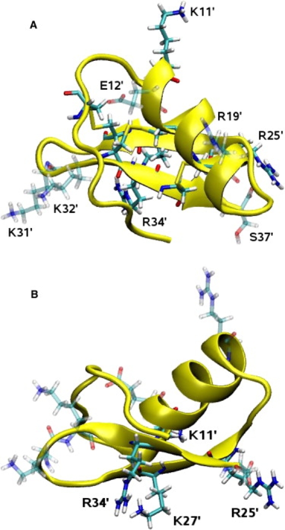
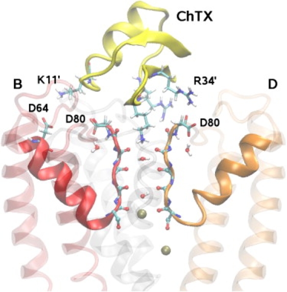
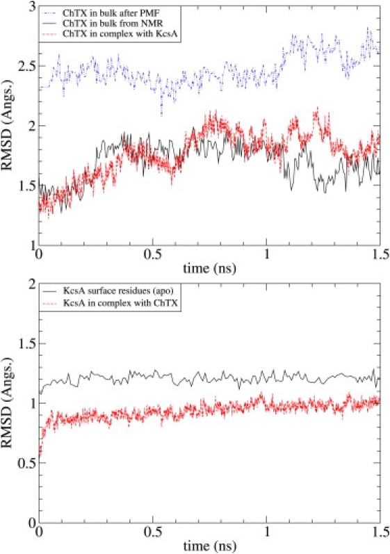
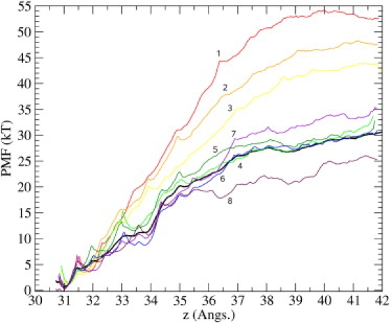
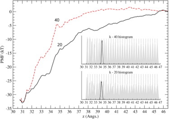
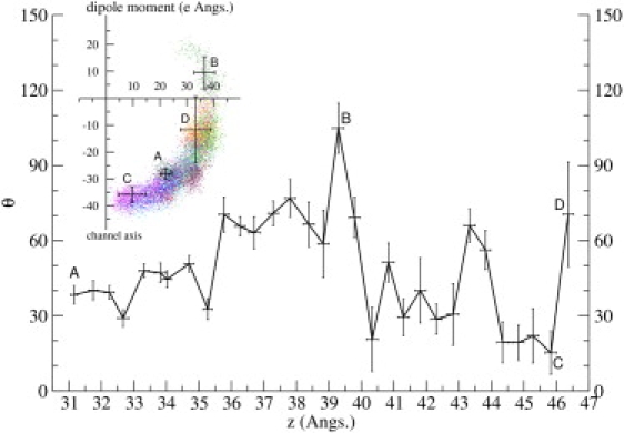
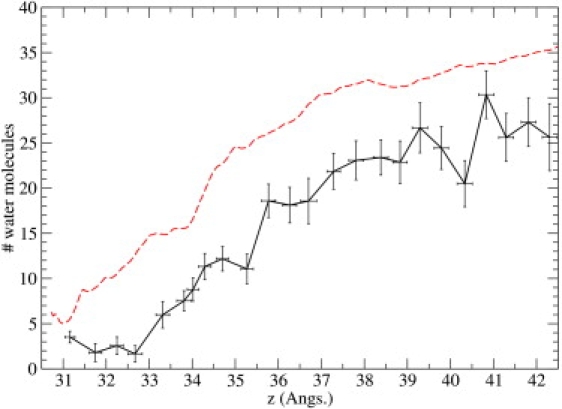
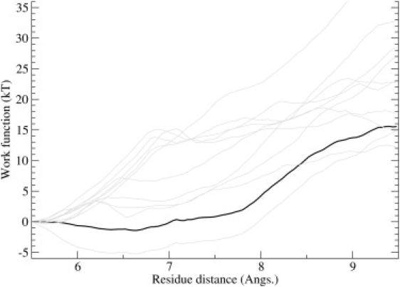
Ion channel-toxin complexes are ideal systems for computational studies of protein-ligand interactions, because, in most cases, the channel axis provides a natural reaction coordinate for unbinding of a ligand and a wealth of physiological data is available to check the computational results. We use a recently determined structure of a potassium channel-charybdotoxin complex in molecular dynamics simulations to investigate the mechanism and energetics of unbinding. Pairs of residues on the channel protein and charybdotoxin that are involved in the binding are identified, and their behavior is traced during umbrella-sampling simulations as charybdotoxin is moved away from the binding site. The potential of mean force for the unbinding of charybdotoxin is constructed from the umbrella sampling simulations using the weighted histogram analysis method, and barriers observed are correlated with specific breaking of interactions and influx of water molecules into the binding site. Charybdotoxin is found to undergo conformational changes as a result of the reaction coordinate choice--a nontrivial decision for larger ligands--which we explore in detail, and for which we propose solutions. Agreement between the calculated and the experimental binding energies is obtained once the energetic consequences of these conformational changes are included in the calculations.


Figures

Figure 1
Figure 1
NMR structure of ChTX from two different perspectives. (A) View from the top, as it is docked in the KcsA channel. The three disulfide bonds that confer some rigidity to ChTX are clearly visible (indicated by yellow). (B) Side view, where the arginine and lysine residues involved in the binding are indicated. Visual figures in this article have been rendered using Tachyon (Tachyon Software, Denver, CO) within the VMD environment.
Figure 2
Figure 2
Side view of ChTX in complex with a KcsA potassium channel surrogate. ChTX backbone is shown in yellow and the side chains of K11′, K27′, R25′, and R34′ residues involved in the binding are explicitly shown. Two of the four monomers in KcsA (B and D) are shown clearly. The monomer A is removed and C is shown as a shadow for clarity. The carbonyl groups in the filter and the side chains of D80 and D64 residues are explicitly indicated. The water molecules in the filter and two K+ ions (one at the S4 binding site and one in the cavity) are also shown.
Figure 3
Figure 3
The RMSD of ChTX in bulk water, in complex with the KcsA channel, and in the last umbrella sampling window (top). The NMR structure 2CRD1 is used as the reference state. RMSD of the surface residues of KcsA (i.e., 49–64 in S5-P and 78–87 in F-S6) with and without ChTX bound (bottom). The NMR structure of the complex 2A9H is used as the reference state.
Figure 4
Figure 4
Distributions of the distances between the strongly interacting pairs in the KcsA channel-ChTX complex. The K27′-Y78 histogram shows the N-O distances for each of the four monomers denoted by AD. Three H atoms in the amide group make hydrogen bonds with the carbonyls of A, B, and D, while that of C is left free. The elongated tail in the R34′-D80 plot stems from equilibration—this interaction is established shortly after system equilibration and remains associated for the rest of the simulation.
Figure 5
Figure 5
Convergence study of the PMF. Data from the first 11 Å is used as a guide to check for sufficient convergence. Here, vertical axis refers to the estimated PMF, and horizontal axis, z, refers to the COM position of the ChTX backbone heavy-atoms, with z = 0 defined as the center of the simulation box (this convention holds throughout the article). The binding site is located at z ∼ 31 Å measured along the channel axis. The last 3.2 ns of the MD data is divided into eight equal parts and a PMF is constructed from each set. The PMFs are sequentially numbered from 1 to 8, as indicated in the figure.
Figure 6
Figure 6
Effect of increasing the number of windows on the PMF. The density data used in Fig. 5 are augmented by including extra windows at 0.25 Å intervals, up to z ∼ 36 Å as shown by the dotted lines. The window number and positions are indicated in the figure. The resulting PMF (solid line), is compared to the first one obtained using 0.5 Å intervals (dashed line).
Figure 7
Figure 7
Comparison of the PMFs obtained from the umbrella sampling simulations using the force constants of 20 kcal/mol/Å2 (solid line) and 40 kcal/mol/Å2 (dashed line). The same routine employed in the construction of the k-20 PMF has been used for the k-40 PMF. (Inset) Distribution of the ChTX-COM as overlapping histograms along the reaction coordinate. For each window, we display the actual distribution of the ChTX-COM (shaded histogram) and its constraint coordinate (dotted line, usually to the right). (Solid curve) Distribution for the extra window (to cover the poorly sampled region, see Methods).
Figure 8
Figure 8
Comparison of the NMR structure of ChTX (light shaded) with that obtained from the last umbrella window (dark shaded). The side chains of the residues Z1′, V16′, and L20′ are explicitly shown. K27′ (in licorice representation) is also displayed to give a sense of orientation. (Dashed lines) The V16′-L20′ hydrogen bond in the last turn of the α-helix, which is broken by tidal forces during pulling.
Figure 9
Figure 9
Average distances of interacting pairs in the KcsA channel and ChTX, plotted as a function of the reaction coordinate z. Additional pair distances are included in the K27′ plot to show its sequential transfer along the channel backbone. The trajectory data are from the k-20 umbrella sampling simulations. Error bars indicate one standard deviation.
Figure 10
Figure 10
Orientation and fluctuations of the dipole moment of ChTX plotted as a function of the reaction coordinate z. The angle θ is defined between the dipole moment and the −z axis (toward the channel). (Inset) Polar graph of the dipole moment sampled from all the k-20 umbrella windows, where individual data points are plotted as a vector with angle θ from the channel axis and radius equal to its dipole magnitude. Error bars indicate one standard deviation.
Figure 11
Figure 11
Number of water molecules in the KcsA channel-ChTX interface plotted as a function of the reaction coordinate z. (Dashed line) Relevant PMF values, purely for comparison purposes. Trajectory data are derived from the k-20 umbrella sampling simulations.
Figure 12
Figure 12
PMF estimate for the conversion of the ChTX structure from the NMR conformer to the distorted conformer. The horizontal axis refers to the COM distance between V16′ backbone and L20′ backbone. Individual work functions are shown in shading, while the PMF obtained from Jarzynski's equation is shown in solid representation.

References

    1. Fersht A. Freeman; New York: 1999. Structure and Mechanism in Protein Science.
    1. Halperin I., Ma B., Wolfson H., Nussinov R. Principles of docking: an overview of search algorithms and a guide to scoring functions. Proteins. 2002;47:409–443. - PubMed
    1. Brooijmans N., Kuntz I.D. Molecular recognition and docking algorithms. Annu. Rev. Biophys. Biomol. Struct. 2003;32:335–373. - PubMed
    1. Gabdoulline R.R., Wade R.C. Brownian dynamics simulation of protein-protein diffusional encounter. Methods. 1998;14:329–341. - PubMed
    1. Wang W., Donini O., Reyes C.M., Kollman P.A. Biomolecular simulations: recent developments in force fields, simulations of enzyme catalysis, protein-ligand, protein-protein, and protein-nucleic acid noncovalent interactions. Annu. Rev. Biophys. Biomol. Struct. 2001;30:211–243. - PubMed

Publication types

LinkOut - more resources