. 2009 Apr 8;96(7):2832-48.
doi: 10.1016/j.bpj.2008.12.3941.

Calmodulin mediates the Ca2+-dependent regulation of Cx44 gap junctions

Affiliations

Calmodulin mediates the Ca2+-dependent regulation of Cx44 gap junctions

Yubin Zhou et al. Biophys J. .

Abstract

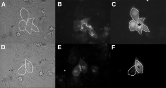
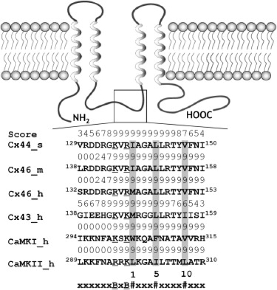
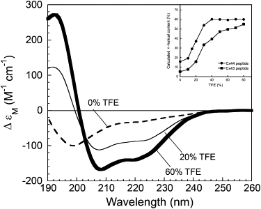
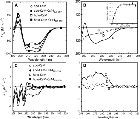
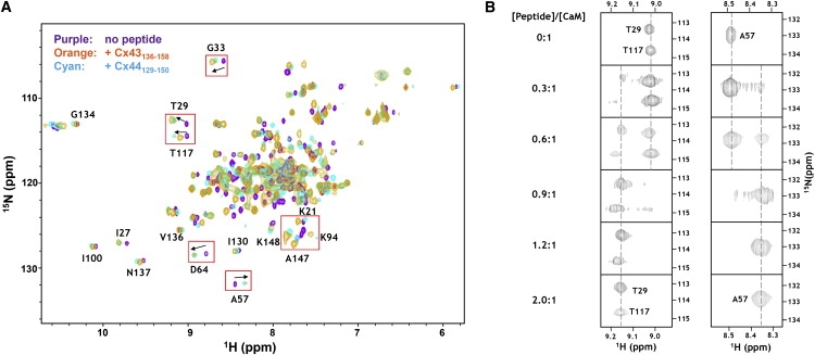
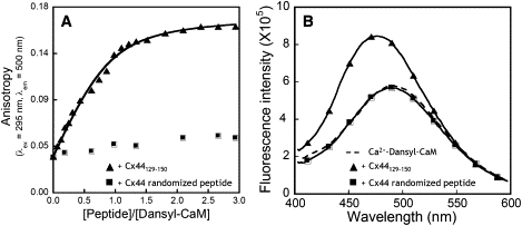
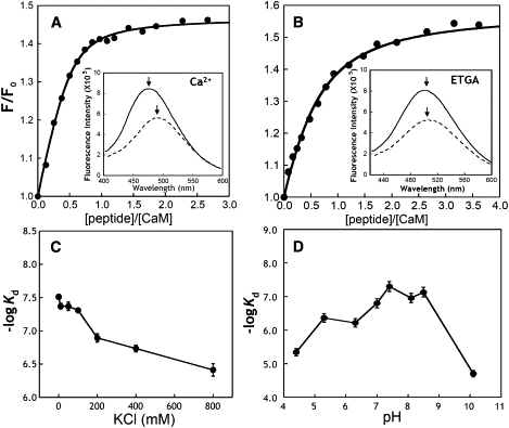
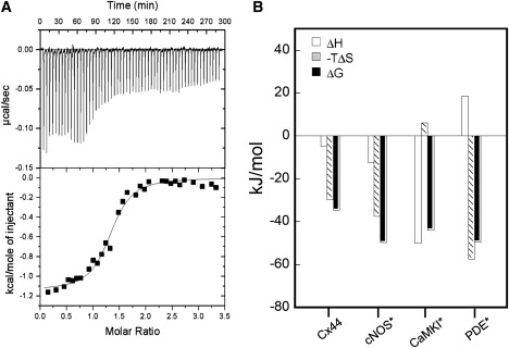
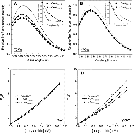
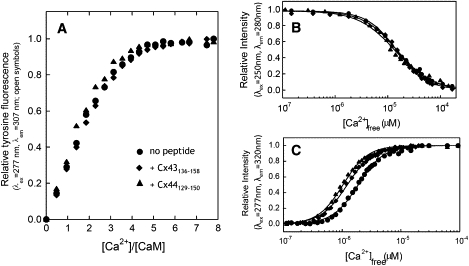
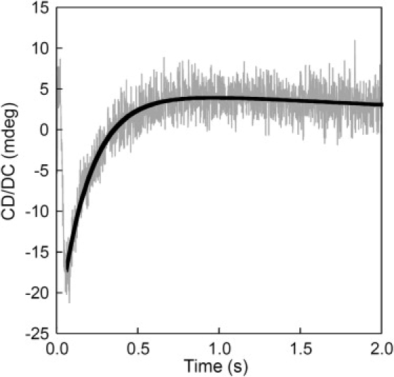
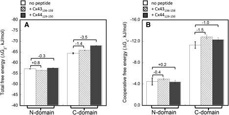
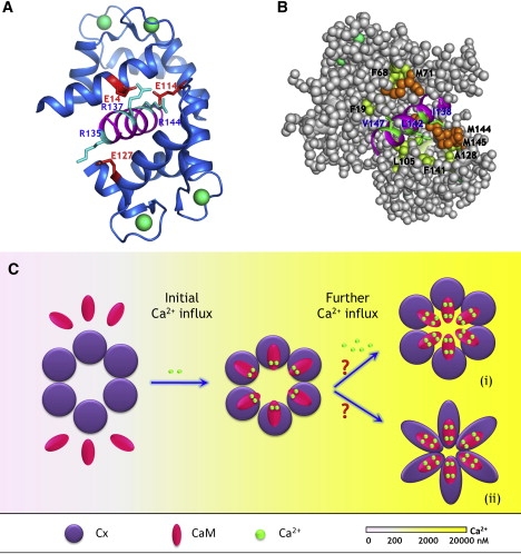
We have shown previously that the Ca2+-dependent inhibition of lens epithelial cell-to-cell communication is mediated in part by the direct association of calmodulin (CaM) with connexin43 (Cx43), the major connexin in these cells. We now show that elevation of [Ca2+](i) in HeLa cells transfected with the lens fiber cell gap junction protein sheep Cx44 also results in the inhibition of cell-to-cell dye transfer. A peptide comprising the putative CaM binding domain (aa 129-150) of the intracellular loop region of this connexin exhibited a high affinity, stoichiometric interaction with Ca2+-CaM. NMR studies indicate that the binding of Cx44 peptide to CaM reflects a classical embracing mode of interaction. The interaction is an exothermic event that is both enthalpically and entropically driven in which electrostatic interactions play an important role. The binding of the Cx44 peptide to CaM increases the CaM intradomain cooperativity and enhances the Ca2+-binding affinities of the C-domain of CaM more than twofold by slowing the rate of Ca2+ release from the complex. Our data suggest a common mechanism by which the Ca2+-dependent inhibition of the alpha-class of gap junction proteins is mediated by the direct association of an intracellular loop region of these proteins with Ca2+-CaM.


Figures

Figure 1
Figure 1
A sustained elevation in [Ca2+]i inhibits Cx44-EYFP cell-to-cell dye transfer. Cx44-EYFP gap junction-mediated biochemical coupling was measured in 80%–90% confluent monolayers of transiently transfected HeLa cells by Lucifer Yellow dye transfer. Representative experimental results for both resting and elevated [Ca2+]i conditions, phase images (A and D) expressed Cx44-EYFP fluorescence (B and E), and LY dye injection results (C and F) are shown. (AC) Resting [Ca2+]i (25 nM). (DF) Elevated [Ca2+]i (1270 nM). The injected cell is denoted by an asterisk ().
Figure 2
Figure 2
Membrane topology and the putative CaM-binding site in α-class connexins including Cx43 and Cx44 (or human Cx46). The integral membrane protein Cx is composed of four transmembrane (TM) segments (helices), two extracellular loops, one cytoplasmic loop, a short N-terminus, and a longer C-terminal tail. The predicted CaM binding sites are located in the second half of the intracellular loop between TM2 and TM3. The numeric score ranges from 1–9, representing the probability of an accurate prediction of a high affinity CaM binding site (18). Similar to the Ca2+/CaM-dependent kinase I (CaMKI) and II (CaMKII), the identified CaM-binding sequences in α-class connexins fits the 1-5-10 subclass, where each number represents the presence of a hydrophobic residue. #, Hydrophobic residues (highlighted in gray); B, basic residues (underscored); m, mouse; s, sheep; h, human.
Figure 3
Figure 3
Far UV CD spectra of the synthetic peptide Cx44129–150 with 0% (dashed line), 20% (thin solid line), and 60% (bold solid line) TFE (v/v). The inset shows calculated α-helical content as a function of TFE concentration for the peptides Cx44129–150 (●) and Cx43136–158 (■).
Figure 4
Figure 4
CD studies of the interaction between the Cx44 peptide and CaM. (A) Far UV circular dichroism spectra of CaM in the presence of 5 mM EGTA (○, apo-CaM) or CaCl2 (□, holo-CaM), and a 1:1 CaM-peptide mixture with 5 mM EGTA (●, apo-CaM-peptide) or CaCl2 (■, holo-CaM-peptide). (B) The far UV CD spectra of Cx44129–150 (dotted line, curve a) and the calculated difference spectrum with 5 mM EGTA (dashed line, curve b) or Ca2+ (solid line, curve c). The inset showed the relative change of CD signals at 222 nm as a function of the synthetic peptide in the presence 5 mM Ca2+. (C) Near UV CD spectra of CaM in the presence of 5 mM EGTA (○, apo-CaM) or CaCl2 (□, holo-CaM), and 1:1 CaM-peptide complex with 5 mM EGTA (●, apo-CaM-peptide) or 5 mM CaCl2 (■, holo-CaM-peptide). (D) The near UV circular dichroism spectra of Cx44129–150 (dotted line, curve a) and the calculated difference spectrum with 5 mM EGTA (dashed line, curve b) or Ca2+ (solid line, curve c). All the spectra were recorded at room temperature in 10 mM Tris, 100 mM KCl, at pH 7.5.
Figure 5
Figure 5
Monitoring the interaction between CaM and the Cx peptides by (1H, 15N)-HSQC spectroscopy. (A) An overlay of HSQC spectra of holo-CaM (purple) with the spectrum of the holo-CaM-Cx44129–150 complex (cyan) or the holo-CaM-Cx43136–158 complex (orange). Representative peaks that exhibited significant movement on peptide binding were framed by boxes. (B) Titration of holo-CaM with Cx44129–150. Note that the progressive disappearance of peaks was accompanied by the concomitant appearance of corresponding peaks at new positions.
Figure 6
Figure 6
The dansyl fluorescence (A) anisotropy and (B) emission spectra of dansylated CaM at increasing concentration of Cx44129–150 (▴) or the Cx44 randomized control peptide (■). Dansyl-CaM with a concentration of 2 μM was titrated with the peptide solutions in a buffer consisting of 50 mM Tris-HCl, 5 mM CaCl2, 100 mM KCl at pH 7.5. The fluorescence anisotropy were measured at λex = 335 nm and λem = 500 nm with an integration time of 20 s.
Figure 7
Figure 7
Interaction of Cx44129–150 with dansyl-CaM monitored by steady-state fluorescence. (A) The titration curve of dansyl-CaM (1.25 μM) with Cx44129–150 in the presence of 5 mM Ca2+ in a salt-free buffer consisting of 50 mM Tris, pH 7.5. The inset showed the fluorescence spectrum of dansyl-CaM in the presence (solid line) or absence (dashed line) of equivalent amount of Cx44129–150. (B) The titration curve of dansyl-CaM with Cx44129–150 in the presence of 5 mM EGTA in a salt-free buffer consisting of 50 mM Tris, pH 7.5. The inset showed the fluorescence spectrum of dansyl-CaM in the presence (solid line) or absence (dashed line) of equivalent amount of Cx44129–150. The emission maxima were indicated by arrows. (C) Plot of −logKd as a function of varying amount of KCl in 5 mM CaCl2, 50 mM Tris, pH 7.5. (D) pH dependence of Cx44129–150 binding to CaM. The binding affinities were derived from the peptide titration curve of dansyl-CaM.
Figure 8
Figure 8
(A) ITC microcalorimetric traces and the derived isotherms of 25 μM CaM titrated with ∼500 μM Cx44129–150 in 20 mM PIPES, 100 mM KCl, 2 mM CaCl2, pH 6.8 at 25°C. (B) Comparison of the enthalpic (ΔH) and entropic (−TΔS) contributions to the Gibbs free energy (ΔG) on the formation of the holo-CaM-peptide complexes. Data from Brokx et al. (36).
Figure 9
Figure 9
Use of CaM mutants to monitor the interaction between CaM and Cx peptides. (A and B) Interaction of the Cx peptides with 5 μM CaM mutants T26W and Y99W. Fluorescence emission was acquired from 325 nm to 415 nm with excitation wavelength at 295 in the absence (solid line) or presence of Cx44129–150 (▴) and Cx43136–158 (♦). The inset showed the normalized signal changes (integrated area from 325 nm to 415 nm) as a function of added Cx peptides in 50 mM Tris, 5 mM Ca Cl2, 100 mM KCl at pH 7.5. (C and D) Acrylamide quenching of the tryptophan fluorescence in CaM mutants T26W and Y99W in the absence (○) or presence of Cx44129–150 (▴) or Cx43136–158 (♦).
Figure 10
Figure 10
Ca2+ titration of CaM with and without the bound Cx peptides. (A) Stoichiometric Ca2+ titration of CaM (●), and CaM bound to the peptide Cx44129–150 (▴) or Cx43136–158 (♦) in 50 mM Tris, 100 mM KCl at pH 7.5. (B and C) Equilibrium Ca2+ titration of CaM (●) and CaM in complex with Cx44129–150 (▴) or Cx43136–158 (♦) in 100 mM KCl, 50 mM HEPES, pH 7.5. Domain-specific (B) intrinsic phenylalanine (λex = 250 nm, λem = 280 nm) or (C) tyrosine fluorescence (λex = 277 nm, λem = 320 nm) was monitored to report the equilibrium Ca2+-binding constants of N- or C-domain of CaM, respectively. The Ca2+ indicator dye Oregon Green 488 BAPTA-5N was used to measure the ionized Ca2+ concentration. All the experiments were repeated at least in triplicate.
Figure 11
Figure 11
Stopped-flow trace for the EGTA-induced dissociation of Ca2+ from the CaM-Cx44129–150 complex. Syringe 1 contained 2 μM CaM-peptide (1:2) mixture with 0.1 mM Ca and syringe 2 consisted of equal volume of 10 mM EGTA.
Figure 12
Figure 12
Comparison of the total free energies ΔG2 (A) and the intradomain cooperative free energies ΔGc (B) of Ca2+ binding to each domain of CaM with and without the Cx peptides. The energies were obtained from the equilibrium titration curves shown in Fig. 10, B and C, according to Eqs. 4 and 5. ΔG2 standards for the total free energy for the binding of two Ca2+ ions to either N- or C-domain of CaM and accounts for any cooperativity between the two sites in each domain. By assuming that both sites in each domain have equal intrinsic Ca2+-binding affinities, the lower limit for changes in free energy due to cooperative Ca2+ binding (ΔGc) was calculated according to Eq. 5. The changes in free energies after and before Cx peptides binding were indicated. All the experiments were repeated for at least three trials.
Figure 13
Figure 13
Proposed model of Ca2+-mediated regulation of gap junction permeability. (A and B) Modeled structure of the CaM-Cx44129–150 complex. The model structure is built based on the 3D structure of holo-CaM in complex with the CaM binding region from CaMKII (pdb entry: 1cdm) using MODELER. Residues involved in potential (A) electrostatic interactions and (B) hydrophobic interactions were indicated. Specifically, residues R135, R137, and R144 from the peptide (cyan) are capable of forming salt bridges with residues E127, E14, and E114 (red) from holo-CaM, respectively. In addition, hydrophobic residues from the peptide (V147, V142, and I138, green) is in close proximity with F19/F68/M71, and L105/F141, and A128/M144/M145 from holo-CaM (Met, orange; Phe and Ala, yellow), respectively. (C) Proposed model of regulation of gap junction inhibition mediated by CaM. The subtle changes in the intracellular Ca2+ concentration (at submicromolar range) are initially sensed by the “high-affinity” C-domain of CaM, enabling it to preferably interact with the intracellular loop of Cx proteins. Such interaction enhances the efficiency and sensitivity of intracellular Ca2+ sensing because of increases in both Ca2+-binding affinity and intradomain cooperativity within the C-domain of CaM. The partially saturated, Cx-bound CaM might serve as an intermediate state to prevent the free diffusion of CaM in the cytoplasm. Once the intracellular Ca2+ is further elevated to micromolar range, the half-saturated CaM is then able to quickly respond to the Ca2+ signals and triggers the fully open conformation that is capable of leading to the inhibition of intercellular communication mediated by gap junction. The entity responsible for the Ca2+-mediated inhibition of gap junction still remains to be defined. It could arise from the physical obstruction of the pore by holo-CaM (state i), or be due to the holo-CaM triggered conformational changes in Cx protein itself (state ii). Whether or not all six subunits have to bind calmodulin to exhibit Ca2+-dependent gap junction inhibition remains to be defined.

Similar articles

Cited by

References

    1. Mathias R.T., Rae J.L., Baldo G.J. Physiological properties of the normal lens. Physiol. Rev. 1997;77:21–50. - PubMed
    1. Eiberger J., Degen J., Romualdi A., Deutsch U., Willecke K. Connexin genes in the mouse and human genome. Cell Commun. Adhes. 2001;8:163–165. - PubMed
    1. Eastman S.D., Chen T.H., Falk M.M., Mendelson T.C., Iovine M.K. Phylogenetic analysis of three complete gap junction gene families reveals lineage-specific duplications and highly supported gene classes. Genomics. 2006;87:265–274. - PubMed
    1. Krutovskikh V., Yamasaki H. Connexin gene mutations in human genetic diseases. Mutat. Res. 2000;462:197–207. - PubMed
    1. Gao Y., Spray D.C. Structural changes in lenses of mice lacking the gap junction protein connexin43. Invest. Ophthalmol. Vis. Sci. 1998;39:1198–1209. - PubMed

Publication types

MeSH terms

LinkOut - more resources