TNF activates astrocytes and catecholaminergic neurons in the solitary nucleus: implications for autonomic control
- PMID: 19348788
- PMCID: PMC2693276
- DOI: 10.1016/j.brainres.2009.03.059
TNF activates astrocytes and catecholaminergic neurons in the solitary nucleus: implications for autonomic control
Abstract
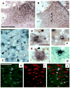
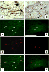
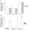
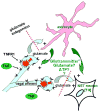
Tumor necrosis factor [TNF] produces a profound anorexia associated with gastrointestinal stasis. Our work suggests that the principal site of action of TNF to cause this change in gastric function is via vagal afferents within the nucleus of the solitary tract [NST]. Excitation of these afferents presumably causes gastric stasis by activating downstream NST neurons that, in turn, suppress gastric motility via action on neurons in the dorsal motor nucleus of the vagus that project to the stomach. Results from our parallel studies on gastric vago-vagal reflexes suggest that noradrenergic neurons in the NST are particularly important to the generation of reflex gastroinhibition. Convergence of these observations led us to hypothesize that TNF action in the NST may preferentially affect putative noradrenergic neurons. The current study confirms our observations of a dose-dependent TNF activation of cells [as indicated by cFOS production] in the NST. The phenotypic identity of these TNF-activated neurons in the NST was approximately 29% tyrosine hydroxylase [TH]-positive [i.e., presumably noradrenergic neurons]. In contrast, less than 10% of the nitrergic neurons were activated after TNF exposure. Surprisingly, another 54% of the cFOS-activated cells in the NST were phenotypically identified to be astrocytes. Taken together with previous observations, the present results suggest that intense or prolonged vagal afferent activity [induced by visceral pathway activity, action of gut hormones or cytokines such as TNF] can alter local astrocyte immediate early gene expression that, in turn, can provoke long-term, perhaps permanent changes in the sensitivity of vagal-reflex circuitry.
Figures


Similar articles
-
Involvement of glutamate in gastrointestinal vago-vagal reflexes initiated by gastrointestinal distention in the rat.Auton Neurosci. 2003 Jan 31;103(1-2):19-37. doi: 10.1016/s1566-0702(02)00145-5. Auton Neurosci. 2003. PMID: 12531396
-
CXCL12 sensitizes vago-vagal reflex neurons in the dorsal medulla.Brain Res. 2013 Jan 25;1492:46-52. doi: 10.1016/j.brainres.2012.11.022. Epub 2012 Nov 23. Brain Res. 2013. PMID: 23178697 Free PMC article.
-
TNFalpha-stimulation of cFos-activation of neurons in the solitary nucleus is suppressed by TNFR:Fc adsorbant construct in the dorsal vagal complex.Brain Res. 2003 Jun 20;976(1):69-74. doi: 10.1016/s0006-8993(03)02687-8. Brain Res. 2003. PMID: 12763623
-
TNFalpha: a trigger of autonomic dysfunction.Neuroscientist. 2008 Feb;14(1):53-67. doi: 10.1177/1073858407305725. Epub 2007 Oct 2. Neuroscientist. 2008. PMID: 17911224 Free PMC article. Review.
-
TNF(alpha) modulation of visceral and spinal sensory processing.Curr Pharm Des. 2005;11(11):1391-409. doi: 10.2174/1381612053507828. Curr Pharm Des. 2005. PMID: 15853670 Review.
Cited by
-
Astrocytes in the Ventromedial Hypothalamus Involve Chronic Stress-Induced Anxiety and Bone Loss in Mice.Neural Plast. 2021 Jul 8;2021:7806370. doi: 10.1155/2021/7806370. eCollection 2021. Neural Plast. 2021. PMID: 34306063 Free PMC article.
-
PAR1-activated astrocytes in the nucleus of the solitary tract stimulate adjacent neurons via NMDA receptors.J Neurosci. 2015 Jan 14;35(2):776-85. doi: 10.1523/JNEUROSCI.3105-14.2015. J Neurosci. 2015. PMID: 25589770 Free PMC article.
-
Astrocytes in the nucleus of the solitary tract: Contributions to neural circuits controlling physiology.Physiol Behav. 2020 Sep 1;223:112982. doi: 10.1016/j.physbeh.2020.112982. Epub 2020 Jun 11. Physiol Behav. 2020. PMID: 32535136 Free PMC article. Review.
-
Participation of α2 -adrenoceptors in sodium appetite inhibition during sickness behaviour following administration of lipopolysaccharide.J Physiol. 2016 Mar 15;594(6):1607-16. doi: 10.1113/JP270377. Epub 2015 Jul 7. J Physiol. 2016. PMID: 26036817 Free PMC article. Review.
-
Thrombin action on astrocytes in the hindbrain of the rat disrupts glycemic and respiratory control.Am J Physiol Regul Integr Comp Physiol. 2020 Jun 1;318(6):R1068-R1077. doi: 10.1152/ajpregu.00033.2020. Epub 2020 Apr 22. Am J Physiol Regul Integr Comp Physiol. 2020. PMID: 32320636 Free PMC article.
References
-
- Andrews PL, Davis CJ, Bingham S, Davidson HI, Hawthorn J, Maskell L. The abdominal visceral innervation and the emetic reflex: pathways, pharmacology, and plasticity. Can J Physiol Pharmacol. 1990;68:325–45. - PubMed
-
- Araque A, Parpura V, Sanzgiri RP, Haydon PG. Tripartite synapses: glia, the unacknowledged partner. Trends Neurosci. 1999;22:208–15. - PubMed
-
- Arenander A, de Vellis J. Early response gene induction in astrocytes as a mechanism for encoding and integrating neuronal signals. Prog Brain Res. 1992;94:177–88. - PubMed
-
- Barros VG, Duhalde-Vega M, Caltana L, Brusco A, Antonelli MC. Astrocyte-neuron vulnerability to prenatal stress in the adult rat brain. J Neurosci Res. 2006;83:787–800. - PubMed
Publication types
MeSH terms
Substances
Grants and funding
LinkOut - more resources
Full Text Sources

