. 2009 Jun 19;284(25):16872-16881.
doi: 10.1074/jbc.M809348200. Epub 2009 Apr 6.

Latent transforming growth factor beta-binding proteins and fibulins compete for fibrillin-1 and exhibit exquisite specificities in binding sites

Affiliations

Latent transforming growth factor beta-binding proteins and fibulins compete for fibrillin-1 and exhibit exquisite specificities in binding sites

Robert N Ono et al. J Biol Chem. .

Abstract

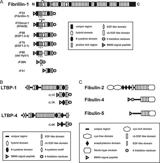
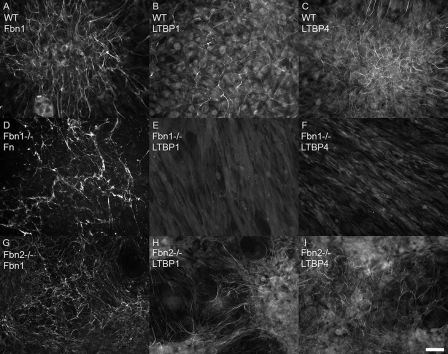
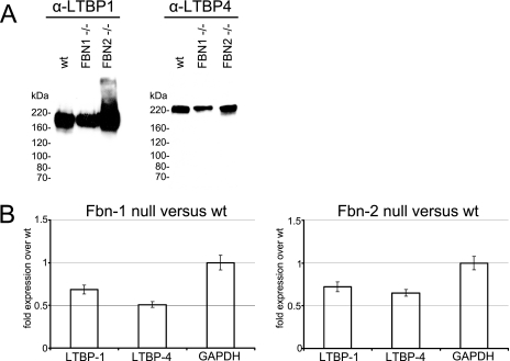
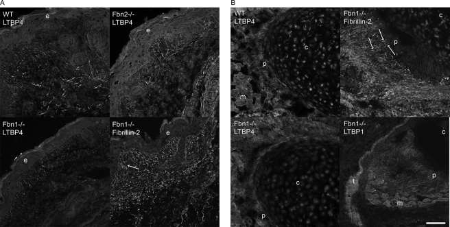
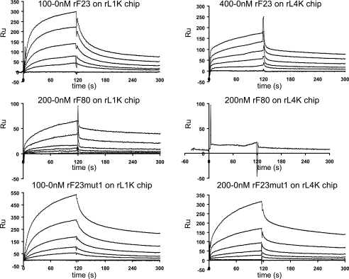
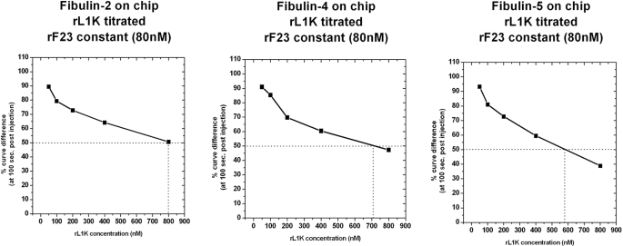
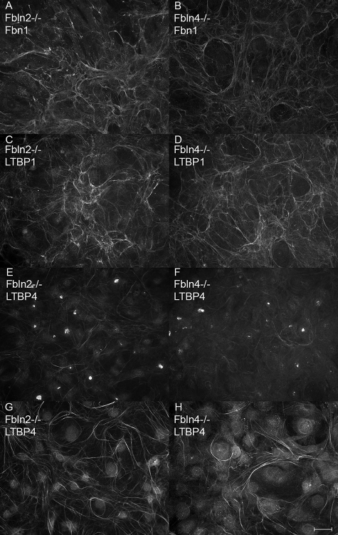
Latent transforming growth factor (TGF) beta-binding proteins (LTBPs) interact with fibrillin-1. This interaction is important for proper sequestration and extracellular control of TGFbeta. Surface plasmon resonance interaction studies show that residues within the first hybrid domain (Hyb1) of fibrillin-1 contribute to interactions with LTBP-1 and LTBP-4. Modulation of binding affinities by fibrillin-1 polypeptides in which residues in the third epidermal growth factor-like domain (EGF3) are mutated demonstrates that the binding sites for LTBP-1 and LTBP-4 are different and suggests that EGF3 may also contribute residues to the binding site for LTBP-4. In addition, fibulin-2, fibulin-4, and fibulin-5 bind to residues contained within EGF3/Hyb1, but mutated polypeptides again indicate differences in their binding sites in fibrillin-1. Results demonstrate that these protein-protein interactions exhibit "exquisite specificities," a phrase commonly used to describe monoclonal antibody interactions. Despite these differences, interactions between LTBP-1 and fibrillin-1 compete for interactions between fibrillin-1 and these fibulins. All of these proteins have been immunolocalized to microfibrils. However, in fibrillin-1 (Fbn1) null fibroblast cultures, LTBP-1 and LTBP-4 are not incorporated into microfibrils. In contrast, in fibulin-2 (Fbln2) null or fibulin-4 (Fbln4) null cultures, fibrillin-1, LTBP-1, and LTBP-4 are incorporated into microfibrils. These data show for the first time that fibrillin-1, but not fibulin-2 or fibulin-4, is required for appropriate matrix assembly of LTBPs. These studies also suggest that the fibulins may affect matrix sequestration of LTBPs, because in vitro interactions between these proteins are competitive.


Figures

FIGURE 1.
FIGURE 1.
Schematic diagrams of the fibrillin-1 recombinant polypeptides (A), LTBP recombinant polypeptides (B), and fibulin recombinant proteins (C) used in this study. Domain modules contained within each peptide are depicted. In A, EGF3 and Hyb1 are bracketed at the top of the diagram. Exact amino acid sequences of the mutated peptides (rF23mut1, rF68, rF79, and rF80) are shown in supplemental Table S2.
FIGURE 2.
FIGURE 2.
Matrix deposition of fibrillin-1, LTBP-1, and LTBP-4 by fibroblast cultures. Immunofluorescence, using antibodies specific for fibrillin-1 (pAb 9543) (A and G), LTBP-1 (pAb 8579) (B, E, and H), and LTBP-4 (pAb 2101) (C, F, and I), demonstrated incorporation of all three proteins into fibrillar matrices in wild type fibroblasts (A–C) grown for 9 days. In Fbn1 null fibroblasts (D–F) grown for 13 days, no matrix incorporation of LTBPs was visible (E and F), even though fibronectin fibrils were present in the Fbn1 null matrix (D). Fbn2 null fibroblasts (G–I), grown for 9 days, assemble fibrillin-1 positive fibrils (G) as well as LTBP-1 (H) and LTBP-4 (I) positive fibrils. Bar = 20 μm.
FIGURE 3.
FIGURE 3.
A, Western blot analyses of medium proteins from wild type, Fbn1 null, and Fbn2 null fibroblasts. Equal numbers of cells from the different cell lines were aliquoted into wells in serum-containing medium. The next day serum-containing medium was replaced with serum-free medium. Serum-free medium was collected after 2 more days in culture. Total medium proteins were precipitated, applied to 5% SDS-PAGE, transferred to nitrocellulose, and blotted with antibodies specific for LTBP-1 and LTBP-4. Results show that wild type (wt), Fbn1 null, and Fbn2 null fibroblasts secrete LTBP-1 and LTBP-4 into the medium. B, quantitative real-time PCR from Fbn1 null fibroblasts and Fbn2 null fibroblasts compared with wild type littermate fibroblasts. Expression levels of LTBP-1 and LTBP-4 were comparable in Fbn1 null and Fbn2 null fibroblasts. The experiments were conducted in triplicate. Average values from these experiments are shown, with error bars. Glyceraldehyde-3-phosphate dehydrogenase (GAPDH) was quantitated as a control.
FIGURE 4.
FIGURE 4.
Immunofluorescence of tissues from wild type, Fbn1 null, and Fbn2 null mice. A, P0 skin showed equivalent patterns of pAb 2101 LTBP-4 positive fibrils in both wild type and Fbn2 null sections. Fibrillin-2 staining was used to identify positive microfibrillar staining in Fbn1 null skin; characteristic fibrillin fibrils perpendicular to the dermal-epidermal junction are indicated (arrow), and typical thicker and longer fibrils can be seen in the deeper dermis. LTBP-4 staining patterns in skin were very similar to these fibrillin patterns at the dermal-epidermal junction and in the deeper dermis. In contrast to wild type and Fbn2 null skin sections, many fewer LTBP-4 positive fibrils were seen in Fbn1 null sections. B, LTBP-4 staining was also apparently reduced in sections of perichondrium from Fbn1 null compared with wild type mice. Fibrillin-2 staining showed positive fibrils (arrows) in the perichondrium of Fbn1 null mice. pAb 8579 LTBP-1 staining of perichondrium and tendon in Fbn1 null sections were fibrillar and similar to fibrillin staining patterns. e, epidermis; c, cartilage; p, perichondrium; m, muscle; t, tendon. Bar, 50 μm.
FIGURE 5.
FIGURE 5.
BiaCore sensorgrams of titrations of rF23, rF80, and rF23mut1 on LTBP-1- and LTBP-4-coated chips. Each experiment was performed as a series of titrated analytes in solution, flowed over a chip coupled with ligand. Titrations are indicated above each set of representative binding curves. Values listed in Table 1 were obtained from experiments similar to the ones shown in this figure. In some cases, titrations were performed multiple times. RU, resonance units.
FIGURE 6.
FIGURE 6.
Competition between LTBP-1 and fibrillin-1 for fibulins. Fibulin-2, fibulin-4, and fibulin-5 were each used to coat a BiaCore chip. A constant amount (80 nm) of recombinant fibrillin-1 (rF23) was preincubated with increasing amounts (50–800 nm) of LTBP-1 competitor (rL1K). Binding of rF23 without competitor is set as 100%, and results with competitor LTBP-1 are shown as a percentage of this maximal binding. Titrations of LTBP-1 show that LTBP-1 binding to fibrillin-1 competes with fibrillin-1 binding to fibulin-2, fibulin-4, and fibulin-5.
FIGURE 7.
FIGURE 7.
Matrix deposition of fibrillin-1, LTBP-1 and LTBP-4 in fibulin-2 and fibulin-4 null cultures. Immunofluorescence, using antibodies specific for fibrillin-1 (pAb 9543) (A and B), LTBP-1 (pAb 8579) (C and D), and LTBP-4 (pAb 2101) (E–H), demonstrated incorporation of all three proteins into fibrillar matrices in both fibulin-2 null (A, C, E, and G) and fibulin-4 null (B, D, F, and H) cells. Cells were grown for 4 (A–F) and 9 days (G and H). Bar, 20 μm.

References

    1. Charbonneau N. L., Ono R. N., Corson G. M., Keene D. R., Sakai L. Y. ( 2004) Birth Defects Res. C Embryo Today 72, 37– 50 - PubMed
    1. Sengle G., Charbonneau N. L., Ono R. N., Sakai L. Y. ( 2008) Primer on the Metabolic Bone Diseases and Disorders of Mineral Metabolism, 7th Edition, pp. 27– 32, American Society for Bone and Mineral Research, Washington, DC
    1. Sakai L. Y., Keene D. R., Engvall E. ( 1986) J. Cell Biol. 103, 2499– 2509 - PMC - PubMed
    1. Zhang H., Hu W., Ramirez F. ( 1995) J. Cell Biol. 129, 1165– 1176 - PMC - PubMed
    1. Charbonneau N. L., Dzamba B. J., Ono R. N., Keene D. R., Corson G. M., Reinhardt D. P., Sakai L. Y. ( 2003) J. Biol. Chem. 278, 2740– 2749 - PubMed

Publication types

MeSH terms