Review
. 2009 May;174(5):1575-87.
doi: 10.2353/ajpath.2009.080987. Epub 2009 Apr 6.

Surveillance of antigen-presenting cells by CD4+ CD25+ regulatory T cells in autoimmunity: immunopathogenesis and therapeutic implications

Affiliations
Review

Surveillance of antigen-presenting cells by CD4+ CD25+ regulatory T cells in autoimmunity: immunopathogenesis and therapeutic implications

Sébastien André et al. Am J Pathol. 2009 May.

Abstract

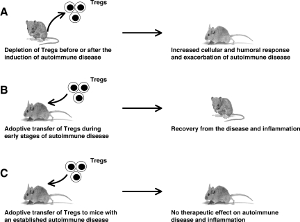
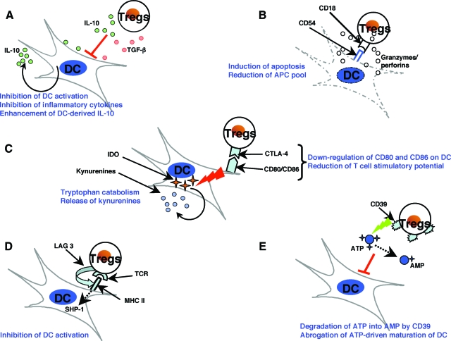
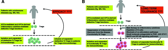
CD4+CD25+ regulatory T cells (Tregs) play a critical role in preventing immune aggression. One way in which Tregs exert immune surveillance activities is by modifying the function of antigen presenting cells (APCs) such as dendritic cells, macrophages, and B cells. Tregs can induce apoptosis of APCs or inhibit their activation and function, thereby regulating subsequent innate and adaptive immune responses. These actions of Tregs are mediated by both soluble factors (interleukin [IL]-10, transforming growth factor-beta, perforins, granzymes) and cell-associated molecules (cytotoxic T lymphocyte antigen 4, lymphocyte activation gene-3, CD18, neuropilin-1, LFA-1/CD11a, CD39), of which cytotoxic T lymphocyte antigen 4 has a key role. However, in autoimmunity, chronically activated APCs under the influence of intracellular signaling pathways, such as phosphatidyl inositol 3 kinase, JAK-STAT, MAPK, and nuclear factor-kappaB pathways, can escape surveillance by Tregs, leading to the activation of T cells that are refractory to suppression by Tregs. Moreover, APCs and APC-derived inflammatory cytokines such as tumor necrosis factor, IL-6, IL-1beta, and IL-23 can render Tregs defective and can also reciprocally enhance the activity of the IL-17-producing pathogenic Th17 T cell subset. Emerging knowledge of the importance of APC-Treg interactions in maintaining immune tolerance and aberrations in this cross talk in autoimmune diseases provides a rationale for therapeutic approaches specifically targeting this axis of the immune system.


Figures

Figure 1
Figure 1
Role of Tregs in the pathogenesis of autoimmune diseases: lessons from experimental models. A: Using several experimental animal models, it has been demonstrated that depletion of Tregs in animals before or after the induction of autoimmune disease leads to increased cellular and humoral responses and to an exacerbation of disease. B: Adoptive transfer of Tregs at the time of induction of disease has been shown to decrease the severity of disease and inflammation, suggesting that Tregs have a potential therapeutic application in the early stages of an autoimmune disease. C: Transfer of Tregs appears unable to cure established chronic inflammatory process, in part due to the influence of chronically activated APCs.
Figure 2
Figure 2
The mechanisms of suppression of dendritic cells by Tregs. Several mutually nonexclusive mechanisms have been proposed to account for the modulation of DCs by Tregs. The suppression of DCs by Tregs is mediated by both soluble factors and cell-associated molecules. However, recent reports suggest that CTLA-4 molecule has a key role in Treg-mediated suppressive functions. A: Treg-derived IL-10 and TGF-β induce a suppressive phenotype in DCs and inhibit inflammatory cytokine expression by DCs while enhancing the secretion of the anti-inflammatory cytokine IL-10. B: Activated Tregs express high levels of granzyme A and perforin and exert CD18/CD54 adhesive interaction-dependent cytotoxicity against both immature and mature DCs, thus reducing the antigen-presenting and co-stimulatory pool. C: CTLA-4 interaction with B7 molecules transduces a negative signal for DCs CTLA-4 diminishes the co-stimulatory molecules CD80 and CD86 and converts DCs into less potent APCs with reduced T cell stimulatory potential. CTLA-4 can also induce indoleamine 2,3-dioxygenase (IDO) expression in DCs. IDO catalyzes the conversion of tryptophan into kynurenines, which are potent immunosuppressive metabolites. D: Tregs can interact with MHC class II on DCs via lymphocyte activation gene-3 (LAG-3), a CD4-related transmembrane protein that inhibits DC activation by a mechanism involving ERK-mediated recruitment of SHP-1. E: Tregs express CD39 (nucleoside triphosphate diphosphohydrolase-1), an ectoenzyme that degrades ATP to AMP. Thus, Tregs reduce ATP-mediated activation of DCs and the adjuvant activities of ATP. Some of these mechanisms are also reported to be critical in Treg-mediated regulation of functions of macrophages and B cells.
Figure 3
Figure 3
Neutralization of inflammatory cytokines and blocking APC function are critical for the success of Treg immunotherapy in chronic autoimmune and inflammatory diseases. A: Tregs are reported to be defective in several autoimmune diseases such as multiple sclerosis (MS) and rheumatoid arthritis (RA). Therefore, Treg-immunotherapy, by isolating and expanding these ‘defective’ Tregs, might not be efficacious in inducing tolerance. Further, ongoing inflammation within the host could also render Tregs defective after adoptive transfer. B: In such conditions, therapeutic restoration of the Treg compartment in vivo either by targeting inflammatory cytokines or B cells, blocking co-stimulatory pathways, or targeting intracellular signal transduction pathways (PI3K, JAK-STAT, MAPK, or NF-κB signaling pathways) might constitute a primary goal. Once conditions are established in which Treg function is restored, Treg-immunotherapy by isolating, expanding, and re-infusing these competent cells could be considered as a second step for inducing long-term immune tolerance.

References

    1. Sakaguchi S, Yamaguchi T, Nomura T, Ono M. Regulatory T cells and immune tolerance. Cell. 2008;133:775–787. - PubMed
    1. Tang Q, Bluestone JA. The Foxp3+ regulatory T cell: a jack of all trades, master of regulation. Nat Immunol. 2008;9:239–244. - PMC - PubMed
    1. Bennett CL, Christie J, Ramsdell F, Brunkow ME, Ferguson PJ, Whitesell L, Kelly TE, Saulsbury FT, Chance PF, Ochs HD. The immune dysregulation, polyendocrinopathy, enteropathy X-linked syndrome (IPEX) is caused by mutations of FOXP3. Nat Genet. 2001;27:20–21. - PubMed
    1. Wildin RS, Ramsdell F, Peake J, Faravelli F, Casanova JL, Buist N, Levy-Lahad E, Mazzella M, Goulet O, Perroni L, Bricarelli FD, Byrne G, McEuen M, Proll S, Appleby M, Brunkow ME. X-linked neonatal diabetes mellitus, enteropathy and endocrinopathy syndrome is the human equivalent of mouse scurfy. Nat Genet. 2001;27:18–20. - PubMed
    1. Hori S, Nomura T, Sakaguchi S. Control of regulatory T cell development by the transcription factor Foxp3. Science. 2003;299:1057–1061. - PubMed

Publication types

MeSH terms

Substances