

Sites of differential DNA methylation between placenta and peripheral blood: molecular markers for noninvasive prenatal diagnosis of aneuploidies
- PMID: 19349366
- PMCID: PMC2671250
- DOI: 10.2353/ajpath.2009.081038
Sites of differential DNA methylation between placenta and peripheral blood: molecular markers for noninvasive prenatal diagnosis of aneuploidies
Abstract
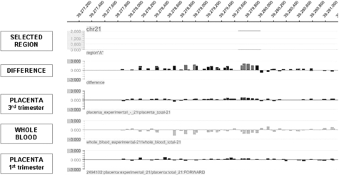
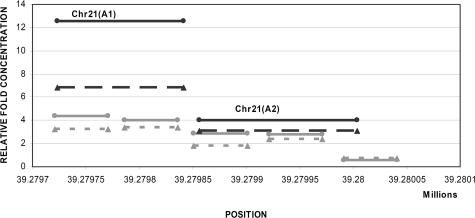
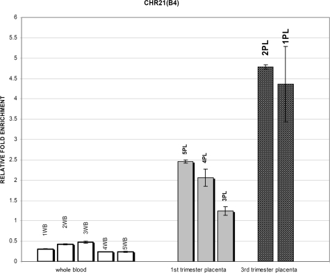
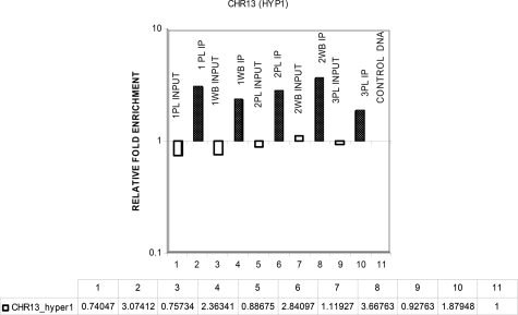
The use of epigenetic differences between maternal whole blood and fetal (placental) DNA is one of the main areas of interest for the development of noninvasive prenatal diagnosis of aneuploidies. However, the lack of detailed chromosome-wide identification of differentially methylated sites has limited the application of this approach. In this study, we describe an analysis of chromosome-wide methylation status using methylation DNA immunoprecipitation coupled with high-resolution tiling oligonucleotide array analysis specific for chromosomes 21, 18, 13, X, and Y using female whole blood and placental DNA. We identified more than 2000 regions of differential methylation between female whole blood and placental DNA on each of the chromosomes tested. A subset of the differentially methylated regions identified was validated by real-time quantitative polymerase chain reaction. Additionally, correlation of these regions with CpG islands, genes, and promoter regions was investigated. Between 56 to 83% of the regions were located within nongenic regions whereas only 1 to 11% of the regions overlapped with CpG islands; of these, up to 65% were found in promoter regions. In summary, we identified a large number of previously unreported fetal epigenetic molecular markers that have the potential to be developed into targets for noninvasive prenatal diagnosis of trisomy 21 and other common aneuploidies. In addition, we demonstrated the effectiveness of the methylation DNA immunoprecipitation approach in the enrichment of hypermethylated fetal DNA.
Figures
References
-
- Hultén MA, Dhanjal S, Pertl B. Rapid and simple prenatal diagnosis of common chromosome disorders advantages and disadvantages of the molecular methods FISH and QF-PCR. Reproduction. 2003;126:279–297. - PubMed
-
- Lo YM, Corbetta N, Chamberlain PF, Rai V, Sargent IL, Redman CW, Wainscoat JS. Presence of fetal DNA in maternal plasma and serum. Lancet. 1997;350:485–487. - PubMed
-
- Lo YM, Hjelm NM, Fidler C, Sargent IL, Murphy MF, Chamberlain PF, Poon PM, Redman CW, Wainscoat JS. Prenatal diagnosis of fetal RhD status by molecular analysis of maternal plasma. N Engl J Med. 1998;339:1734–1738. - PubMed
-
- Bianchi DW, Avent ND, Costa JM, Van der Schoot CE. Noninvasive prenatal diagnosis of fetal Rhesus D: ready for Prime(r) Time. Obstet Gynecol. 2005;106:841–844. - PubMed
Publication types
MeSH terms
Substances
LinkOut - more resources
Full Text Sources
Other Literature Sources
Medical
Molecular Biology Databases
