. 2009 Sep;136(3):926-933.
doi: 10.1378/chest.09-0336. Epub 2009 Apr 6.

Utility of [18F]2-fluoro-2-deoxyglucose-PET in sporadic and tuberous sclerosis-associated lymphangioleiomyomatosis

Affiliations

Utility of [18F]2-fluoro-2-deoxyglucose-PET in sporadic and tuberous sclerosis-associated lymphangioleiomyomatosis

Lisa R Young et al. Chest. 2009 Sep.

Abstract

Mutations in tuberous sclerosis complex (TSC) genes are associated with dysregulated mammalian target of rapamycin (mTOR)/Akt signaling and unusual neoplasms called perivascular epithelioid cell tumors (PEComas), including angiomyolipomas (AMLs) and lymphangioleiomyomatosis (LAM). Tools that quantify metabolic activity and total body burden of AML and LAM cells would be valuable for the assessment of disease progression and the response to therapy in patients with TSC and LAM. Our hypothesis was that constitutive activation of mTOR in LAM and AML cells would result in increased glucose uptake of [(18)F]2-fluoro-2-deoxyglucose (FDG) on PET scanning, as has been suggested by a single prior case report. After institutional review board approval, FDG-PET scanning was performed in six LAM patients. Six additional LAM patients underwent FDG-PET scanning for clinical evaluation of suspected malignancy. Pleural uptake related to prior therapy was identified in four individuals with a remote history of talc pleurodesis. Focal increased uptake was observed in a supraclavicular lymph node in a patient with Hodgkin lymphoma and in a lung nodule in a patient with a biopsy-documented primary lung adenocarcinoma. In one TSC-LAM patient with a biopsy-documented malignant uterine PEComa, robust uptake was noted in metastatic nodules in the lung but not in the LAM-involved lung parenchyma or the patient's massive abdominal lymphangioleiomyomas. No abnormal uptake was identified in the AMLs or LAM lesions in any patients. This pilot study suggests that FDG-PET scans are negative in patients with benign PEComas and therefore are not likely to be useful for estimating the burden of disease in patients with TSC or LAM, but that FDG-PET scans can be used to identify or exclude other neoplasms in these patients.


Figures

Figure 1
Figure 1
Negative FDG-PET scan findings in renal AMLs. Imaging of subject 2 reveals bilateral, large, amorphous, renal AMLs that expand the kidneys and distort the collecting system (A, arrows), but are negative on the FDG-PET scan (B, arrow [note that tracer is seen in renal collecting system only]). An MRI of subject 8 demonstrates bilateral large renal AMLs (C and D), which were negative on the FDG-PET scan (not shown).
Figure 2
Figure 2
Absence of FDG uptake in a lymphangioleiomyoma. CT imaging findings in subject 10 included a large left chylous effusion (A, arrow), and numerous lung cysts, right lower lobe lung nodules, and a large fluid-filled lymphangioleiomyoma (B, arrow). C: FDG-PET scan shows no uptake in the lymphangioleiomyoma. Compressed lung exhibits low-level uptake superior to the pleural fluid (arrowhead). Normal activity is seen in the displaced right urinary tract and myocardium (dashed arrows). A presumed uterine fibroid tumor (8 mm; arrow) has a maximum SUV of 4.8 (hollow arrow), and clinical follow-up was recommended. No uptake was seen in the lung nodules, which had resolved on a follow-up chest CT scan (not shown).
Figure 3
Figure 3
FDG-PET is negative in the lung parenchyma of LAM patients, but abnormal FDG uptake can be seen in the pleura of patients who have undergone prior talc pleurodesis. A: PET-CT scan from subject 8 reveals typical cystic change in the lung and positive FDG uptake consistent with chronic pleural inflammatory reaction after talc pleurodesis (arrow). B and C: subject 5 underwent talc pleurodesis 13 years prior to this FDG-PET scan, which showed uptake in the right pleura and chest wall (arrows).
Figure 4
Figure 4
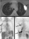
FDG-PET scan identification of a primary lung carcinoma in a patient with LAM. A LAM patient with a primary lung cancer (subject 9) had positive uptake on the FDG-PET scan in the LUL tumor, but not in other LAM disease areas. A PET-CT scan was performed for staging after a needle biopsy revealed a well-differentiated adenocarcinoma in the LUL. The LUL tumor mass (A, white arrow) had an SUV of 3.5 (B and C, arrows). Additionally, there were extensive areas of increased activity throughout the right and left pleura corresponding to nodular pleural thickening (A and B, red arrows). The patient had a history of talc pleurodesis 7 years prior. Follow-up FDG-PET and chest CT scans showed no changes in the pleural abnormalities. The patient had known liver AMLs. No uptake was seen in the liver or in other locations.
Figure 5
Figure 5
Discrimination of benign and malignant PEComas by FDG-PET scan. A TSC-LAM patient with a malignant uterine PEComa and lung nodules had uptake in pulmonary masses but not in abdominal masses or lung tissue on a FDG-PET scan. When this patient (subject 7) was found to have a malignant uterine mass, lung nodules (A, arrow), and large cystic abdominal masses (C, arrows), a widely metastatic uterine sarcoma was suspected. CT/FDG-PET scans performed for staging revealed intense uptake in some, but not all, pulmonary nodules (B and D, arrows) and no uptake in the lung tissue (B) or abdominal cystic lymphangioleiomyomas (C, arrow). Normal activity was present in the myocardium, bladder, and renal collecting system (dashed arrows). Pathology of the uterine lesion revealed marked cellular atypia and an increased number of mitotic figures consistent with a malignant PEComa (hematoxylin-eosin staining, E). Note the diffusely positive staining for desmin (F) and the focal positivity for HMB-45 (G) in a minority of cells. FDG-PET scanning revealed that the abdominal masses were most consistent with benign rather than malignant PEComas, which had important prognostic implications at the time.
Figure 6
Figure 6
Lack of FDG uptake suggests the association of abdominal lymphadenopathy with LAM rather than coincident neoplastic processes. Increased uptake in supraclavicular lymph nodes (A, arrows) was seen in this patient (subject 11) with biopsy-documented Hodgkin disease (nodular sclerosing type). She was presumed to have stage IV disease based on the presence of abdominal masses consistent with lymph nodes with hypodense centers on CT scan that displaced bowel and bladder (black arrow, B). However, these masses were negative on a FDG-PET scan, transcutaneous needle biopsy revealed a spindle cell neoplasm believed to be consistent with LAM, and a high-resolution CT scan of the chest revealed cystic changes that were typical for LAM (not shown). The diagnosis of LAM was made after the patient had completed her course of chemotherapy and radiation therapy. Her staging was revised based on this information. LAM was diagnosed in another subject (subject 12) by video-assisted thoracoscopic surgery lung biopsy after the subject presented with hemoptysis and cystic lung disease. Pelvic lymphadenopathy (C, arrow) was believed to be attributable to LAM, rather than another neoplasm, based on lack of uptake on FDG-PET scan (not shown).

References

    1. Costello LC, Hartman TE, Ryu JH. High frequency of pulmonary lymphangioleiomyomatosis in women with tuberous sclerosis complex. Mayo Clin Proc. 2000;75:591–594. - PubMed
    1. Franz DN, Brody A, Meyer C, et al. Mutational and radiographic analysis of pulmonary disease consistent with lymphangioleiomyomatosis and micronodular pneumocyte hyperplasia in women with tuberous sclerosis. Am J Respir Crit Care Med. 2001;164:661–668. - PubMed
    1. Moss J, Avila NA, Barnes PM, et al. Prevalence and clinical characteristics of lymphangioleiomyomatosis (LAM) in patients with tuberous sclerosis complex. Am J Respir Crit Care Med. 2001;164:669–671. - PubMed
    1. van Slegtenhorst M, de Hoogt R, Hermans C, et al. Identification of the tuberous sclerosis gene TSC1 on chromosome 9q34. Science. 1997;277:805–808. - PubMed
    1. Consortium ECTS. Identification and characterization of the tuberous sclerosis gene on chromosome 16: the European Chromosome 16 Tuberous Sclerosis Consortium. Cell. 1993;75:1305–1315. - PubMed

Substances