. 2009 Apr 13;206(4):893-908.
doi: 10.1084/jem.20082396. Epub 2009 Apr 6.

Impact of a hypomorphic Artemis disease allele on lymphocyte development, DNA end processing, and genome stability

Affiliations

Impact of a hypomorphic Artemis disease allele on lymphocyte development, DNA end processing, and genome stability

Ying Huang et al. J Exp Med. .

Abstract

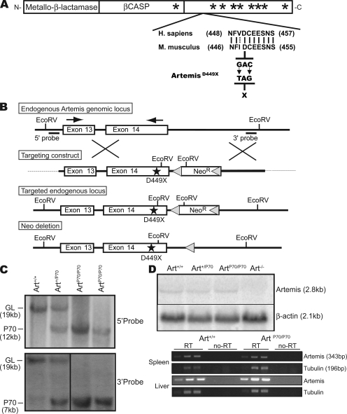
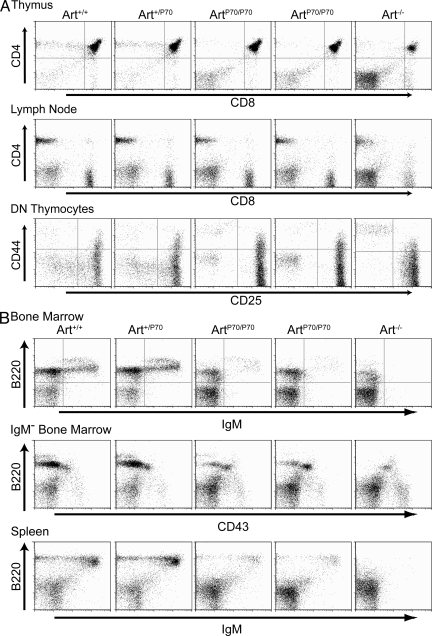
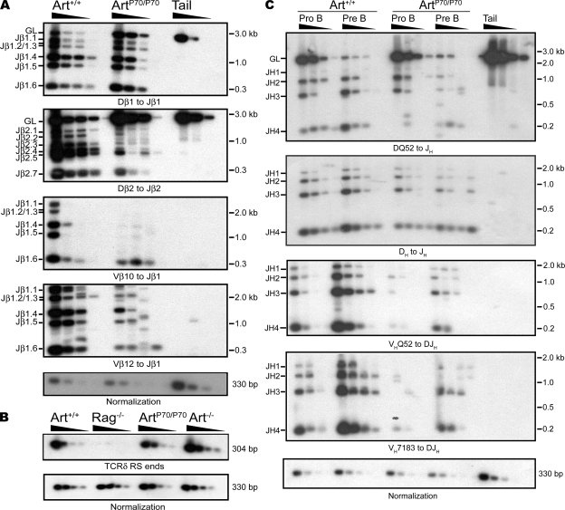
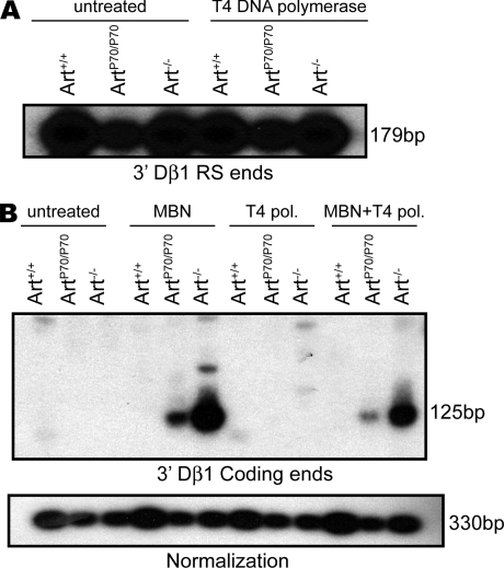
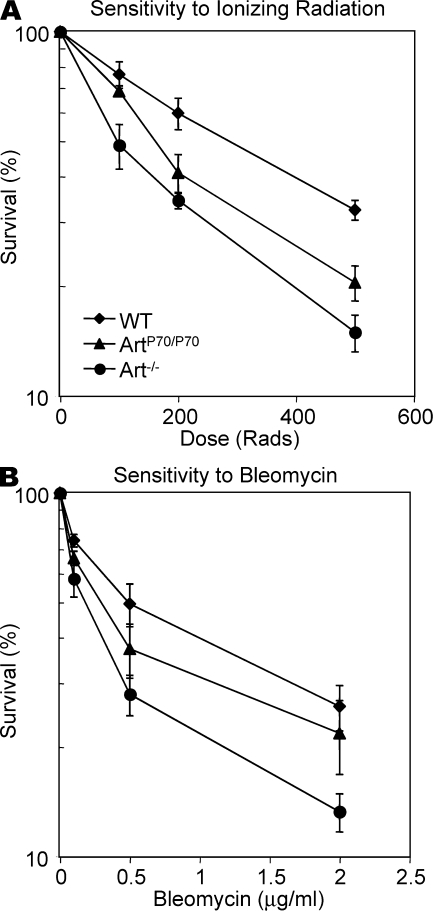
Artemis was initially discovered as the gene inactivated in human radiosensitive T(-)B(-) severe combined immunodeficiency, a syndrome characterized by the absence of B and T lymphocytes and cellular hypersensitivity to ionizing radiation. Hypomorphic Artemis alleles have also been identified in patients and are associated with combined immunodeficiencies of varying severity. We examine the molecular mechanisms underlying a syndrome of partial immunodeficiency caused by a hypomorphic Artemis allele using the mouse as a model system. This mutation, P70, leads to premature translation termination that deletes a large portion of a nonconserved C terminus. We find that homozygous Artemis-P70 mice exhibit reduced numbers of B and T lymphocytes, thereby recapitulating the patient phenotypes. The hypomorphic mutation results in impaired end processing during the lymphoid-specific DNA rearrangement known as V(D)J recombination, defective double-strand break repair, and increased chromosomal instability. Biochemical analyses reveal that the Artemis-P70 mutant protein interacts with the DNA-dependent protein kinase catalytic subunit and retains significant, albeit reduced, exo- and endonuclease activities but does not undergo phosphorylation. Together, our findings indicate that the Artemis C terminus has critical in vivo functions in ensuring efficient V(D)J rearrangements and maintaining genome integrity.


Figures

Figure 1.
Figure 1.
Generation of Artemis-P70 knock-in mice. (A) Hypomorphic Artemis-P70 mutation. Diagram of Artemis cDNA depicting highly conserved metallo-β-lactamase/β-CASP domain (boxes) and nonconserved C terminus with the approximate positions of DNA-PKcs S/Q phosphorylation sites indicated (asterisks). An alignment between the Homo sapiens and Mus musculus amino acid sequences adjacent to the nonsense mutation introduced into the mouse genomic locus is shown. Lines, identical residues; dotted line, conserved residue; arrows, RT-PCR primers. (B) Targeting strategy for the Artemis-P70 knock-in mutation. The endogenous Artemis locus, targeting construct, knock-in allele, and Neo-deleted knock-in allele are depicted. Bars, 5′ and 3′ probes used to screen ES cell clones; stars, Art-P70 mutation. (C) Southern blot analysis of targeted locus. Southern blot analysis of EcoRV-digested genomic DNA from Art+/+, Art+/P70, and ArtP70/P70 kidneys from Neo-deleted mice, hybridized with the 5′ and 3′ probes. Positions of the germline (GL) and targeted (P70) alleles are indicated. (D) Northern blot and RT-PCR analyses of Artemis-P70 mRNA. 20 µg of total RNA isolated from Art+/+, Art+/P70, ArtP70/P70, and Art−/− MEFs was analyzed by Northern blotting using a probe that hybridizes to the 3′ end of the mouse Artemis mRNA. β-Actin was used as a normalization control. RT-PCR was performed on threefold serial dilutions of total RNA from spleen and liver using primers designed to detect transcripts encoding exons 13–14 and a normalization control (tubulin). Each experiment was repeated at least three times with RNA isolated from two independent cell lines and tissues from two mice of each genotype. Representative results are shown. Black lines indicate that intervening lanes have been spliced out.
Figure 2.
Figure 2.
Homozygous Artemis-P70 mice exhibit partial immunodeficiency. Flow cytometric analyses were performed on Art+/+, Art+/P70, ArtP70/P70, and Art−/− mice, as indicated, at 4–5 wk of age. (A) Analysis of T cell development. Thymocytes and lymph node cells were stained with antibodies against the indicated cell surface markers. DN thymocytes were gated for the CD25/CD44 analyses. (B) Analysis of B cell development. Bone marrow and splenocytes were stained with antibodies against the indicated cell surface markers. IgM-negative bone marrow cells (IgM) were gated for B220/CD43 analyses. Five to six mice of each genotype were analyzed. Representative FACS plots are shown.
Figure 3.
Figure 3.
Lymphocyte development defects are caused by impaired V(D)J rearrangements. (A) TCR-β rearrangements. Genomic DNA from purified DN thymocytes from Art+/+ and ArtP70/P70 mice and a nonrearranging tissue (tail) was PCR amplified to detect the indicated Dβ to Jβ and Vβ to DJβ rearrangements. (B) Endogenous TCR-δ signal joints. Levels of extrachromosomal signal joints formed between TCR-Dδ2 and -Jδ1 RSSs were detected by PCR amplification of genomic thymocyte DNA isolated from Art+/+, ArtP70/P70, Art−/−, and RAG2−/− mice. Control PCR amplification of a nonrearranging locus was performed to normalize levels of input DNA. Dδ2-Jδ1 signal joints were detected by Southern blot hybridization using an oligonucleotide probe. (C) IgH rearrangements. Genomic DNA from sorted pro- and pre–B cell populations from Art+/+ and ArtP70/P70 mice and a nonrearranging tissue (tail) was PCR amplified to detect DHQ52 to JH and other DH to JH rearrangements using a degenerate DH primer, as well as VH to DJH rearrangements, as indicated. Experiments in A–C were repeated three times with genomic DNA samples from two different sets of mice. Representative results are shown.
Figure 4.
Figure 4.
V(D)J recombination intermediates in Artemis-P70 thymocytes. (A) Levels of RS ends in Artemis-P70 DN thymocytes. Genomic DNA from sorted DN thymocytes from Art+/+, ArtP70/P70, and Art−/− mice was either untreated or treated with T4 DNA polymerase. Linkers were ligated to the blunt ends, followed by PCR amplification of TCR-Dβ1 3′ RS ends (179 bp). The LM-PCR products were detected by Southern blot hybridization using locus-specific oligonucleotide probes. (B) Accumulation of coding hairpin ends in Artemis-P70 DN thymocytes. The genomic DNA from sorted DN thymocytes was untreated or treated with MBN and/or T4 DNA polymerase, as indicated, and then the LM-PCR reactions were performed to detect 3′ TCR-Dβ1 coding ends (125 bp), as described in A. Control PCR amplification of a nonrearranging locus was performed to normalize levels of input DNA. Experiments in A and B were repeated a minimum of three times with genomic DNA isolated from at least two different mice of each genotype. Representative results are shown.
Figure 5.
Figure 5.
Defective DNA repair and increased genomic instability caused by the Artemis-P70 mutation. (A) IR sensitivity. Art+/+, ArtP70/P70, and Art−/− primary MEFs were exposed to the indicated amounts of IR and then plated in duplicate. The cells were harvested 7 d after IR, stained with trypan blue, and then counted. The percentage of survival compared with untreated cultures is plotted as a function of IR dose. (B) Bleomycin sensitivity. Primary MEFs were plated and then treated with the indicated amounts of bleomycin at 24 h. Cellular survival was determined as described in A. The curves represent the mean of three independent experiments using two independent cell lines of each genotype. Error bars represent SD.
Figure 6.
Figure 6.
Impact of Artemis C-terminal truncations on DNA-PKcs interactions and nuclease activities. (A) Coimmunoprecipitation of Artemis–DNA-PKcs. Constructs expressing c-myc and 6× histidine-tagged full-length (WT), D451X, T432X, and S385X mutant forms of Artemis, and the C terminus alone (C-term), were transfected into 293T cells, immunoprecipitated with α–c-myc, and then analyzed by Western blotting probed with α-6×His antibodies. The membranes were then reprobed with α–DNA-PKcs antibodies. Mock, untransfected 293T cells; (−), no immunoprecipitation; (+), immunoprecipitated. (B) Phosphorylation of Artemis mutants. WT and mutant Artemis proteins (as indicated) purified from 293T cells were incubated with purified DNA-PKcs and γ-[32P]ATP. Phosphorylation products were analyzed by SDS-PAGE followed by autoradiography. Arrowheads, positions of truncated mutant proteins; closed circle, autophosphorylated DNA-PKcs. Black lines indicate that intervening lanes have been spliced out. (C) 5′ to 3′ single-strand exonuclease activity. WT and mutant Artemis proteins (as indicated) were incubated with a 32P 5′ end-labeled single-strand oligonucleotide substrate (20 nt) for 2 h at 37°C. The reactions were analyzed on a 17% denaturing polyacrylamide gel followed by autoradiography. The positions and structures of the input substrate and 1-nt product are indicated on the left. Asterisks, 32P 5′ end label. (D) DNA-PKcs–dependent endonuclease activities in the presence of MgCl2. Endonuclease reactions were performed with a 32P 5′ end-labeled 20-bp hairpin substrate (left) or a 3′ overhang substrate comprised of a [32P] 5′ end-labeled 36-mer annealed to a complementary 21-mer (right) in the presence or absence DNA-PKcs and ATP, as indicated. All reactions contained 10 mM MgCl2 and were analyzed as described in A. The positions and structures of the input substrates and products are indicated. Arrows, sites of endonucleolytic cleavage; asterisks, 32P 5′ end label. (E) DNA-PKcs–independent hairpin opening activity in the presence of MnCl2. Endonuclease reactions were performed with the 32P 5′ end-labeled hairpin and 3′ overhang substrates as described in B, except, the reactions contained 10 mM MnCl2. Reactions were analyzed as described in A. The positions and structures of the input substrates and products are indicated. C, mock transfection; (−), input substrate. All assays were repeated at least three times with proteins from three or more independent transfections. Asterisks, 32P 5′ end label.

References

    1. Bassing C.H., Swat W., Alt F.W. 2002. The mechanism and regulation of chromosomal V(D)J recombination.Cell. 109:S45–S55 - PubMed
    1. Sekiguchi J., Alt F.W., Oettinger M. 2004. The mechanism of V(D)J recombination. Molecular Biology of B cells. Alt F.W., Honjo T., Academic Press, San Diego, CA: 57–78
    1. Fugmann S.D. 2001. RAG1 and RAG2 in V(D)J recombination and transposition.Immunol. Res. 23:23–39 - PubMed
    1. Gellert M. 2002. V(D)J recombination: RAG proteins, repair factors, and regulation.Annu. Rev. Biochem. 71:101–132 - PubMed
    1. Buck D., Malivert L., de Chasseval R., Barraud A., Fondaneche M.C., Sanal O., Plebani A., Stephan J.L., Hufnagel M., le Deist F., et al. 2006. Cernunnos, a novel nonhomologous end-joining factor, is mutated in human immunodeficiency with microcephaly.Cell. 124:287–299 - PubMed

Publication types