. 2009 Apr 13;206(4):937-52.
doi: 10.1084/jem.20082048. Epub 2009 Apr 6.

Alternatively activated macrophage-derived RELM-{alpha} is a negative regulator of type 2 inflammation in the lung

Affiliations

Alternatively activated macrophage-derived RELM-{alpha} is a negative regulator of type 2 inflammation in the lung

Meera G Nair et al. J Exp Med. .

Erratum in

  • J Exp Med. 2009 May 11;206(5):1201

Abstract

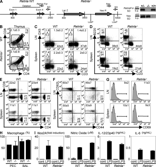
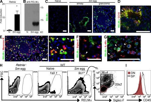
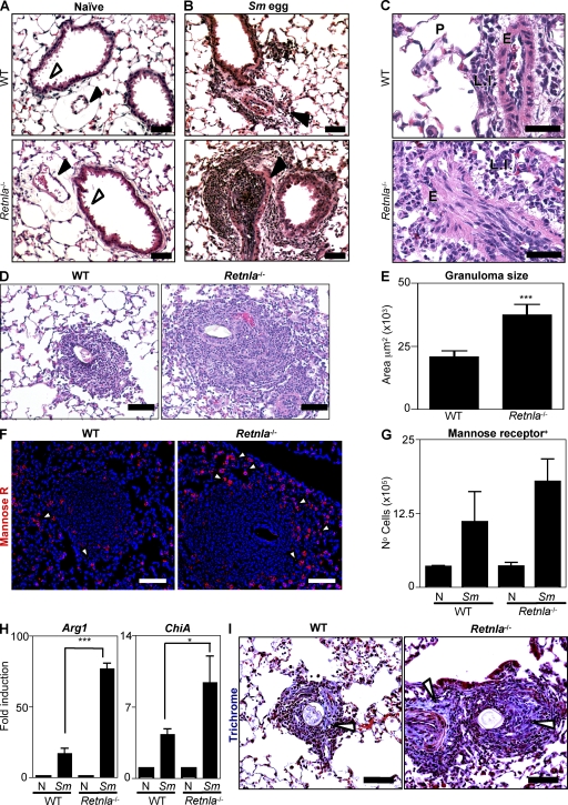
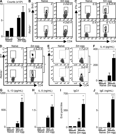
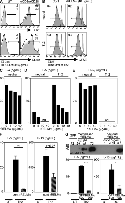
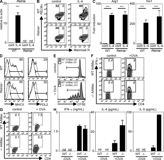
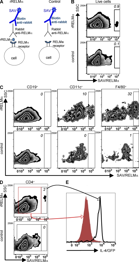
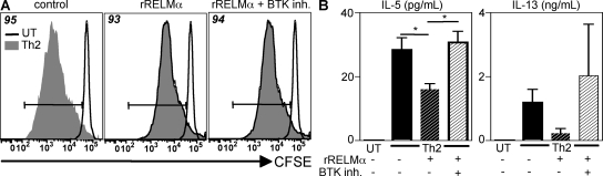
Differentiation and recruitment of alternatively activated macrophages (AAMacs) are hallmarks of several inflammatory conditions associated with infection, allergy, diabetes, and cancer. AAMacs are defined by the expression of Arginase 1, chitinase-like molecules, and resistin-like molecule (RELM) alpha/FIZZ1; however, the influence of these molecules on the development, progression, or resolution of inflammatory diseases is unknown. We describe the generation of RELM-alpha-deficient (Retnla(-/-)) mice and use a model of T helper type 2 (Th2) cytokine-dependent lung inflammation to identify an immunoregulatory role for RELM-alpha. After challenge with Schistosoma mansoni (Sm) eggs, Retnla(-/-) mice developed exacerbated lung inflammation compared with their wild-type counterparts, characterized by excessive pulmonary vascularization, increased size of egg-induced granulomas, and elevated fibrosis. Associated with increased disease severity, Sm egg-challenged Retnla(-/-) mice exhibited elevated expression of pathogen-specific CD4(+) T cell-derived Th2 cytokines. Consistent with immunoregulatory properties, recombinant RELM-alpha could bind to macrophages and effector CD4(+) Th2 cells and inhibited Th2 cytokine production in a Bruton's tyrosine kinase-dependent manner. Additionally, Retnla(-/-) AAMacs promoted exaggerated antigen-specific Th2 cell differentiation. Collectively, these data identify a previously unrecognized role for AAMac-derived RELM-alpha in limiting the pathogenesis of Th2 cytokine-mediated pulmonary inflammation, in part through the regulation of CD4(+) T cell responses.


Figures

Figure 1.
Figure 1.
Generation and characterization of Retnla−/− mice. (A) Schematic diagram of the Retnla locus in WT and Retnla−/− mice and primers used to confirm the genotypes by PCR. (B–G) Frequency of CD4, CD8, B lymphocytes, and NK cells (DX5+) in the thymi, spleen, and LN of naive WT and Retnla−/− mice, and CD4+ T cell surface expression of CD62L, CD44, and CD69. (H) Frequency of resident macrophages from the PEC or the BAL of naive WT and Retnla−/− mice. Results (± SEM of four animals) are representative of two independent experiments of a total number of 9–10 individual animals per group. (I) Real-time PCR analysis of Nos2A expression, and secretion of nitric oxide, IL-12/23p40, and IL-6 by control or LPS-stimulated WT and Retnla−/− macrophages. Results (mean ± SEM) are representative of two independent experiments.
Figure 2.
Figure 2.
RELM-α is expressed after Sm egg challenge. (A) Lung Retnla expression in naive and Sm egg-challenged C57BL/6 mice. *, P <0.05. (B) Western blot analysis of the BAL fluid from naive or day 8 Sm egg-challenged mice. (C) IF staining of lung sections for RELM-α (green) and DAPI (blue). (D–G) Costaining with CC10 (D; red), the mannose receptor (E; red), siglec-F (F; red), or IL-4Rα (G; red). Arrowheads indicate costaining of antibodies with RELM-α. (H) Flow cytometry plots (forward scatter [FSC] vs. RELM-α) of lung cells from naive and Sm egg-challenged mice, and mannose receptor and siglec-F expression by RELM-α–positive cells. *, P <0.05. (I) CD45 expression by the mannose receptorsiglec-F population (DN; red) and mannose receptor+ or siglec-F+ cells (single positive [SP]). Bars, 50 µm. Results (± SEM of three to four animals) are representative of two to three independent experiments (A–H; n = 9–12) and one experiment (I; n = 4).
Figure 3.
Figure 3.
Retnla−/− mice exhibit exacerbated Sm egg-induced pulmonary inflammation. (A–C) H&E-stained lung sections from naive and Sm egg-challenged WT and Retnla−/− mice. Black arrowheads, pulmonary arteries; white arrowheads, bronchiole development. P, lung parenchyma; LI, leukocyte infiltrate; E, endothelium. (D) H&E-stained sections of granulomas from Sm egg-challenged WT and Retnla−/− mice. (E) Size of granulomas surrounding Sm eggs from WT and Retnla−/− mice; n = 30 granulomas from a total of three mice per group. Results are representative of at least two independent experiments with three to four mice per group (n = 6–8 animals per genotype). (F) IF staining of granulomas for the mannose receptor (red) and DAPI (blue). White arrowheads depict mannose receptor+ cells. (G) Flow cytometric quantification of mannose receptor+ cells from dissociated lung tissue. (H) Lung Arg1 and ChiA expression in naive and Sm egg-challenged WT and Retnla−/− mice. ***, P <0.001; *, P <0.05. (I) Masson's trichrome-stained granulomas from WT and Retnla−/− mice. White arrowheads, collagen stain. Bars, 50 µm. Results (mean ± SEM of three to four mice) are representative of two to three independent experiments (n = 6–10 per group).
Figure 4.
Figure 4.
Exacerbated expression of Th2 cytokines in Sm egg-challenged Retnla−/− mice. (A) Cell counts from the draining LN of naive or Sm egg-challenged WT and Retnla−/− mice. (B) Flow cytometric analysis of CD4+ T cell incorporation of BrdU. (C–E) Ex vivo flow cytometric analysis of CD4+ T cell–derived IFN-γ (C), IL-13 (D), and IL-5 (E). (F–H) Antigen-specific secretion of IL-4 (F), IL-13 (G), and IL-5 (H) by draining LN cells. (I) Antigen-specific IgG1 antibody titers. (J) Serum IgE levels. ***, P < 0.001; **, P < 0.01; *, P < 0.05. Results (mean ± SEM of two to four mice per group) are representative of three independent experiments (naive, n = 6; Sm egg-challenged, n = 11).
Figure 5.
Figure 5.
RELM-α negatively regulates Th2 cytokine production by α-CD3/α-CD28–stimulated splenocytes. (A) Expression of CD25 and CD69 by CD4+ T cells from untreated (UT) or α-CD3/α-CD28–stimulated splenocytes in the presence of rRELM-α (filled histogram). (B) Frequency of CFSE-dim CD4+ T cells from untreated splenocytes or splenocytes stimulated with α-CD3/α-CD28 (filled histogram) under neutral or Th2-permissive conditions in the presence rRELM-α. (C–E) Supernatants were analyzed for IL-4 (C), IL-5 (D), and IFN-γ (E) secretion. (F) C3H/HeJ splenocytes were untreated or stimulated with α-CD3/α-CD28 under Th2-permissive conditions in the presence of 5 µg/ml rRELM-α, followed by measurement of IL-5 and IL-13 secretion. (G) RELM-α Western blotting of supernatants (sup) from 293T cells transfected with a control eGFP plasmid or the Retnla plasmid. WT splenocytes were untreated or stimulated with α-CD3/α-CD28 under Th2-permissive conditions in the presence of 72-h sup at a 1:1 ratio followed by measurement of IL-5 and IL-13 secretion. Results (mean ± SEM; ***, P < 0.001; *, P < 0.05) are representative of two to three independent experiments.
Figure 6.
Figure 6.
Retnla−/− AAMacs promote exaggerated production of Th2 cytokines by CD4+ T cells. (A and B) Retnla expression by WT or Retnla−/− UT (cont) or AAMacs was measured by real-time PCR (fold induction over actin; A) and intracellular flow cytometry (B). (C) Arg1 and Ym1 expression (fold induction over cont) was measured. ***, P <0.001. (D) Surface expression of MHC class II and PDL2 by WT and Retnla−/− cont (dashed line) or AAMac (solid line). (E–H) Control or OVA-pulsed WT or Retnla−/− AAMacs were cocultured with CFSE-labeled OVA-specific CD4+ T cells. (E) Frequency of CFSE-dim CD4+ T cells (italics). (F and G) Frequency of IFN-γ+ and IL-4+ CFSE-dim CD4+ T cells (italics). (H) Supernatants were recovered for measurement of IFN-γ, IL-4, and IL-5 secretion. Results (mean ± SEM) are representative of two independent experiments.
Figure 7.
Figure 7.
RELM-α binds to effector CD4+ Th2 cells, DCs, and F4/80+ macrophages. DO11-10/4get splenocytes were incubated with OVA protein, followed by recovery of cells at day 3 to determine which cell types bind RELM-α. (A) Schematic diagram of the RELM-α capture assay. (B) Flow cytometry plot (side scatter [SSC] vs. RELM-α/streptavidin [SAV]) showing frequency of live cells that bind rRELM-α. (C) Frequency of F4/80+ macrophages (right), CD11c+ DCs (middle), and CD19+ B cells (left) that bind RELM-α. (D) Frequency of CD4+ T cells that bind RELM-α (top). (E) IL-4/GFP expression of RELM-α–bound CD4+ T cells (black line) and RELM-α–unbound CD4+ T cells (red histogram). Results are representative of three independent experiments.
Figure 8.
Figure 8.
Suppression of Th2 cytokine production by RELM-α is dependent on BTK signaling. CFSE-labeled splenocytes were left untreated (UT) or stimulated with α-CD3/α-CD28 under Th2-permissive conditions, with or without (+/−) 5 µg/ml rRELM-α, or with or without an inhibitor for BTK (BTK inh.). (A) Frequency of CD4+ T cell proliferation at day 4. (B) IL-5 and IL-13 secretion by cells from A. Results (mean ± SEM; *, P < 0.05) are representative of two independent experiments.

References

    1. Janeway C.A., Jr., Medzhitov R. 2002. Innate immune recognition.Annu. Rev. Immunol. 20:197–216 - PubMed
    1. Stuart L.M., Ezekowitz R.A. 2008. Phagocytosis and comparative innate immunity: learning on the fly.Nat. Rev. Immunol. 8:131–141 - PubMed
    1. Lawrence T., Bebien M., Liu G.Y., Nizet V., Karin M. 2005. IKKalpha limits macrophage NF-kappaB activation and contributes to the resolution of inflammation.Nature. 434:1138–1143 - PubMed
    1. Qualls J.E., Kaplan A.M., van Rooijen N., Cohen D.A. 2006. Suppression of experimental colitis by intestinal mononuclear phagocytes.J. Leukoc. Biol. 80:802–815 - PubMed
    1. Swirski F.K., Libby P., Aikawa E., Alcaide P., Luscinskas F.W., Weissleder R., Pittet M.J. 2007. Ly-6Chi monocytes dominate hypercholesterolemia-associated monocytosis and give rise to macrophages in atheromata.J. Clin. Invest. 117:195–205 - PMC - PubMed

Publication types

MeSH terms