. 2009 May;119(5):1264-74.
doi: 10.1172/JCI36000.

Focal and segmental glomerulosclerosis induced in mice lacking decay-accelerating factor in T cells

Affiliations

Focal and segmental glomerulosclerosis induced in mice lacking decay-accelerating factor in T cells

Lihua Bao et al. J Clin Invest. 2009 May.

Abstract

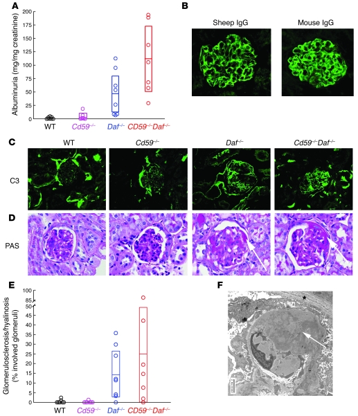
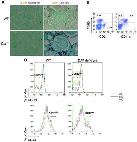
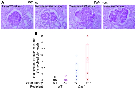
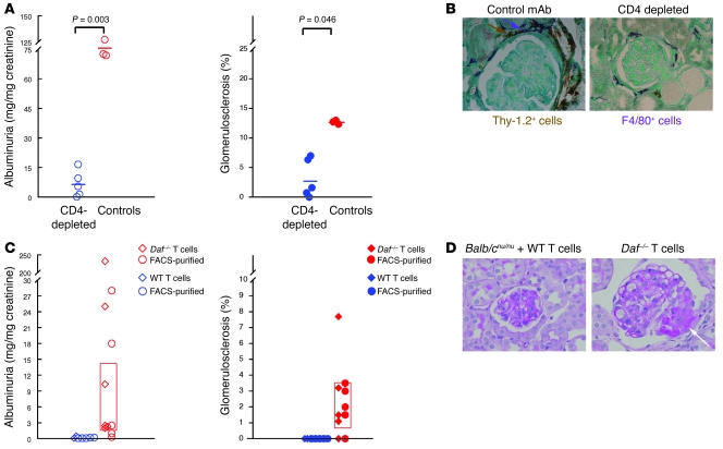
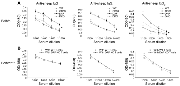
Heritable and acquired diseases of podocytes can result in focal and segmental glomerulosclerosis (FSGS). We modeled FSGS by passively transferring mouse podocyte-specific sheep Abs into BALB/c mice. BALB/c mice deficient in the key complement regulator, decay-accelerating factor (DAF), but not WT or CD59-deficient BALB/c mice developed histological and ultrastructural features of FSGS, marked albuminuria, periglomerular monocytic and T cell inflammation, and enhanced T cell reactivity to sheep IgG. All of these findings, which are characteristic of FSGS, were substantially reduced by depleting CD4+ T cells from Daf(-/-) mice. Furthermore, WT kidneys transplanted into Daf(-/-) recipients and kidneys of DAF-sufficient but T cell-deficient Balb/(cnu/nu) mice reconstituted with Daf(-/-) T cells developed FSGS. In contrast, DAF-deficient kidneys in WT hosts and Balb/(cnu/nu) mice reconstituted with DAF-sufficient T cells did not develop FSGS. Thus, we have described what we believe to be a novel mouse model of FSGS attributable to DAF-deficient T cell immune responses. These findings add to growing evidence that complement-derived signals shape T cell responses, since T cells that recognize sheep Abs bound to podocytes can lead to cellular injury and development of FSGS.


Figures

Figure 1
Figure 1. Anti-podo Ab–induced FSGS in DAF-deficient animals.
(A) Significant albuminuria occurred in Daf–/– and Cd59–/–Daf–/– mice 30 days after administration of anti-podo Abs. (BD) Representative photomicrographs 30 days after anti-podo injection show glomerular localization of sheep and mouse IgG (B) and mouse C3 (C) in the 4 groups; (D) PAS-stained kidney sections show segmental sclerosis (arrows) only in Daf–/– and Cd59–/–Daf–/– mice. (E) The extent of glomerulosclerosis in individual mice 30 days after administration of anti-podo Abs is shown. (F) Extensive podocyte foot process effacement and prominent remodeling of the external surface of the glomerular basement membrane (arrow) was seen by electron microscopy in DAF-deficient mice 30 days after anti-podo Abs administration. Asterisk denotes Bowman capsule. Numeric data shown in A and E from Daf–/– and Daf–/–Cd59–/– mice were normally distributed, with means and 95% CIs shown as horizontal lines and open boxes, respectively. Data from WT and Cd59–/– mice were nonparametric (Anderson-Darling test; P < 0.05); when sufficiently different from the origin, the 95% CI for the median is shown as an open box. Original magnification, ×400 (BD); ×7,100 (F).
Figure 2
Figure 2. Immunological events induced by anti-podo Abs in DAF-deficient mice.
(A) In the kidneys of all mice, there was little neutrophil (brown) or B cell (purple) infiltration, while in DAF-deficient mice, there was marked infiltration with Thy-1.2+ T cells (brown) and F4/80+ monocytic cells (purple). Original magnification, ×400. (B) Flow cytometric analysis of cells isolated from kidneys of DAF-deficient mice 30 days after anti-podo Ab administration showing relative proportions of F4/80+ and CD3+ cells (left panel). The majority of F4/80+ cells were also CD11c+ (right panel). Numbers shown are the percentage of total cells in individual quadrants. (C) Splenic CD4+ cells from WT and DAF-deficient mice were assessed for CD62L (upper panels) and CD44 expression (lower panels) at baseline (d0) and 10 (d10) and 20 days (d20) after passive administration of anti-podo IgG. The CD62Llo and CD44hi populations are denoted by horizontal lines.
Figure 3
Figure 3. Kidney transplantation studies reveal extrarenal DAF deficiency is necessary for development of anti-podo Ab–induced FSGS.
(A) Representative glomerular histopathology 30 days after anti-podo Ab administration in native WT and transplanted Daf–/– kidneys in a WT mouse and native Daf–/– and transplanted WT kidneys in a Daf–/– mouse. FSGS only developed in native and transplanted WT kidneys in Daf–/– mice. Arrows depict regions of segmental sclerosis/hyalinosis. Original magnification, ×400. (B) Shown is the extent of glomerulosclerosis in individual kidneys within each group. Data from native and transplanted kidneys in Daf–/– mice were normally distributed, while native and transplanted kidney data in WT mice were nonparametric (Anderson-Darling test; P < 0.05). Means and 95% CIs are shown as horizontal lines and open boxes, respectively.
Figure 4
Figure 4. Susceptibility to anti-podo Ab–induced FSGS occurs when DAF is absent from T cells.
(A) Shown are urinary albumin excretion (left) and the extent of glomerulosclerosis (right) 30 days after administration of anti-podo Abs in individual Daf–/– mice depleted of CD4+ cells, with mAb GK1.5 or controls receiving irrelevant rat IgG2b. All data were normally distributed (Anderson-Darling test; P > 0.05) and compared by t testing, with P values provided above the data. (B) Infiltration with Thy-1.2+ T cells (brown, arrow) and F4/80+ monocytic cells (purple, arrow) was significantly reduced 30 days after administration of anti-podo Abs in CD4+ cell–depleted but not control Daf–/– mice. (C) Balb/cnu/nu mice reconstituted with DAF-deficient but not WT T cells developed albuminuria and FSGS. Urinary albumin excretion (left) and the percentage of segmentally sclerotic glomeruli (right) in individual Balb/cnu/nu mice that received T cells from WT (blue symbols) or Daf–/– mice (red symbols) 30 days after administration of anti-podo Abs. Experiments differed by whether mice were reconstituted with partially (diamonds) or highly (circles) purified T cells. All data sets in mice receiving T cells from Daf–/– mice were normally distributed with no differences comparing partially and highly purified T cells. Means and 95% CIs for the combined groups are shown as horizontal bars and open boxes, respectively. (D) Representative PAS-stained kidney sections from individual mice show the presence of segmental sclerosis (arrow) only in mice with DAF-deficient T cells. Original magnification, ×600.
Figure 5
Figure 5. Abnormal anti-sheep IgG responses occur when DAF is absent outside T cells.
(A) Serum mouse IgG, IgG1, and IgG2a anti-sheep IgG titers were higher in WT (open diamonds) and Cd59–/– mice (filled squares) compared with Daf–/– (filled circles) and Cd59–/–Daf–/– (X’s) mice. *P < 0.01, IgG concentration in DAF-sufficient versus DAF-deficient mice. (B) Balb/cnu/nu mice reconstituted with T cells from Daf–/– (filled diamonds) or WT mice (open squares) had comparable serum titers of mouse IgG, IgG1, and IgG2a anti-sheep IgG. Data shown represent mean ± SEM.

References

    1. Walport M.J. Advances in immunology: complement (first of two parts). N. Engl. J. Med. 2001;344:1058–1066. doi: 10.1056/NEJM200104053441406. - DOI - PubMed
    1. Speth, C., Prodinger, W.M., Würzner, R., Stoibert, H., and Dierich, M.P. 1993. Complement. In Fundamental immunology. 6th edition. W.E. Paul, editor. Lippincott Williams & Wilkins. Baltimore, Maryland, USA. 1047–1078.
    1. Holers, V.M. 2001. Complement deficiencies. In Clinical immunology: principles and practice. 2nd edition. R. Rich, editor. Mosby. St. Louis, Missouri, USA. 36.1–36.10.
    1. Harris C.L., Rushmere N.K., Morgan B.P. Molecular and functional analysis of mouse decay accelerating factor (CD55). Biochem. J. 1999;341:821–829. doi: 10.1042/0264-6021:3410821. - DOI - PMC - PubMed
    1. Lin F., Emancipator S.N., Salant D.J., Medof M.E. Decay-accelerating factor confers protection against complement- mediated podocyte injury in acute nephrotoxic nephritis. Lab. Invest. 2002;82:563–569. doi: 10.1038/labinvest.3780451. - DOI - PubMed

Publication types

MeSH terms