. 2009 May;11(5):569-79.
doi: 10.1038/ncb1863. Epub 2009 Apr 6.

Border-cell migration requires integration of spatial and temporal signals by the BTB protein Abrupt

Affiliations

Border-cell migration requires integration of spatial and temporal signals by the BTB protein Abrupt

Anna C-C Jang et al. Nat Cell Biol. 2009 May.

Abstract

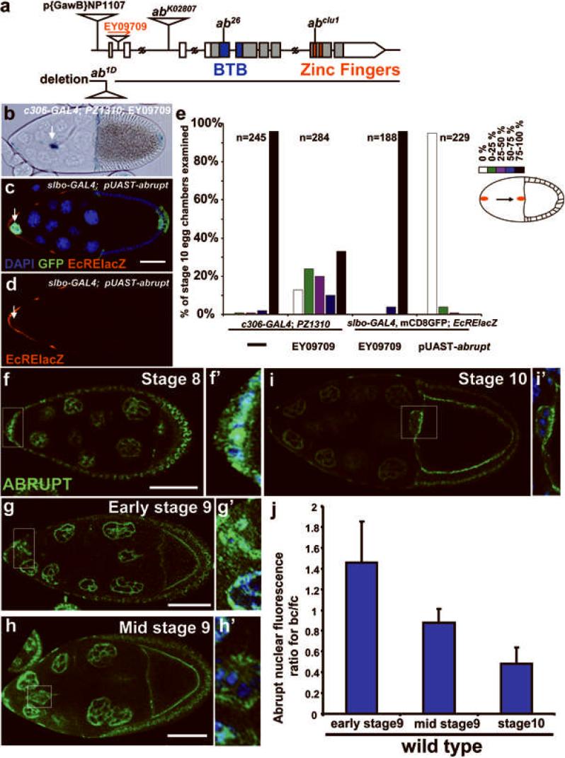
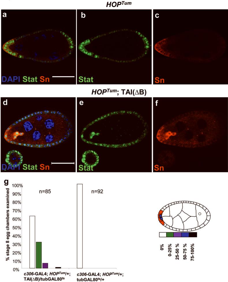
During development, elaborate patterns of cell differentiation and movement must occur in the correct locations and at the proper times. Developmental timing has been studied less than spatial pattern formation, and the mechanisms integrating the two are poorly understood. Border-cell migration in the Drosophila ovary occurs specifically at stage 9. Timing of the migration is regulated by the steroid hormone ecdysone, whereas spatial patterning of the migratory population requires localized activity of the JAK-STAT pathway. Ecdysone signalling is patterned spatially as well as temporally, although the mechanisms are not well understood. In stage 9 egg chambers, ecdysone signalling is highest in anterior follicle cells including the border cells. We identify the gene abrupt as a repressor of ecdysone signalling and border-cell migration. Abrupt protein is normally lost from border-cell nuclei during stage 9, in response to JAK-STAT activity. This contributes to the spatial pattern of the ecdysone response. Abrupt attenuates ecdysone signalling by means of a direct interaction with the basic helix-loop-helix (bHLH) domain of the P160 ecdysone receptor coactivator Taiman (Tai). Taken together, these findings provide a molecular mechanism by which spatial and temporal cues are integrated.


Figures

Figure 1
Figure 1. Spatial patterns of ecdysone signaling in stage 8-9 egg chambers
a-c) Confocal micrographs of egg chambers of the indicated stages triple labeled with DAPI (blue) to stain all nuclei, GFP (green) to show the slbo-GAL4 expression pattern and anti-beta-galactosidase antibodies (red) to show the pattern of expression of the ecdysone reporter EcRE-lacZ. 15 Nurse cells (nc) and one oocyte (o) are surrounded by follicle cells to form a egg chamber. Arrows indicate border cells. Filled arrowheads indicate posterior follicle cells and open arrowheads indicate nurse-cell-associated follicle cells. d-f) the same micrographs as those shown in a-c showing the anti-beta-galactosidase staining only. g) Surface view to show nurse cell associated follicle cell expression of EcRE-lacZ in wild-type. h-i) Effect of dominant-negative EcR (EcRDN) on EcRE-lacZ expression in stage 10 egg chambers. h) Expression of EcRDN with slbo-GAL4 and mCD8-GFP (green) specifically reduced border cell (arrow) but not nurse cell associated follicle cell (open arrowheads) expression of beta-galactosidase (red). i) Same egg chamber as h, showing red channel only. j-l) Expression of a second ecdysone reporter (hs-GAL4-USP;UAS-mCD8-GFP) in stages 8-9 egg chambers. Scale bars represent 50 μm.
Figure 2
Figure 2. Normal and ectopic expression of ecdysone receptor isoforms
a-d) Immunofluorescence staining for the indicated ecdysone receptor isoforms. Follicle cells (open arrowheads), border cells (arrows) and nurse cells (open arrows) are indicated. e) Quantification of the indicated ecdysone receptor subunits. The ratio of immunofluorescence to DAPI staining was measured for 123 border cells and 123 posterior follicle cells at early stage 9. The ratio of border cell (bc) to follicle cell (fc) signal is shown. The error bars represent the standard deviation (n=5-7). f-i) Effects of over-expression of EcRA on EcRE-lacZ expression (red) in clones of follicle cells (green) generated using FLP-OUT GAL4 (see methods for details). f, g) Border cells and h, i) nurse cell associated follicle cells over-expressing EcRA exhibit reduced EcRE-lacZ expression (arrows) compared to neighboring wild-type cells (open arrowheads). j, k) stage 10 egg chamber from a slbo-GAL4; UAS-GFP; EcRE-lacZ female. l, m) Down-regulation of EcRA expression via RNAi. slbo-GAL4; UAS-GFP; UAS-EcRA-dsRNA results in elevated EcRE-lacZ, which is particularly evident in posterior follicle cells (arrow) which normally express little or no detectable β-Galactosidase (arrow in k). n, o) Over-expression of EcRB1 using slbo-GAL4 elevates EcRE-lacZ in posterior follicle cells (compare to k). While all cells that show ectopic EcRE-lacZ also express some GFP, we consistently observed higher levels of EcRE-lacZ in cells expressing low levels of GFP. Scale bars = 50 μm.
Figure 3
Figure 3. Abrupt represses ecdysone signaling in Drosophila egg chambers
a) Genomic organization of the abrupt (ab) locus and domain structure of Abrupt protein. b) Stage 10 egg chamber showing border cell migration defect caused by Abrupt over-expression using c306-GAL4. PZ1310 is a slbo enhancer trap line used to mark border cells (arrow) and centripetal follicle cells. c, d) Stage 10 egg chamber of the genotype slbo-GAL4, mCD8GFP/+; UAS-ab/EcRE-lacZ stained with anti-beta-galactosidase antibodies (red) and DAPI (blue). GFP indicates the slbo-GAL4 expression domain. EcRE-lacZ staining was reduced in border cells (arrow). e) Quantification of border cell migration in the indicated genotypes. The migration path was divided into five sections as shown in the schematic drawing on the right side. The indicated number (n) of stage 10 egg chambers were evaluated for the extent of border cell migration. White bars represent the percentage of egg chambers in which border cells failed to detach from the anterior of the egg chamber. Black bars represent the percentage of egg chambers in which border cells migrated all the way to the oocyte. Other colors represent intermediate phenotypes. f-i) Anti-Abrupt antibody staining (green). f'-i') Higher magnification of the boxed regions shown in (f-i) including DAPI (blue) to mark all nuclei. Scale bars = 50 μm. j) The fluorescence intensity of anti-Abrupt antibody staining was measure for 42 border cells (bc) and 44 oocyte-associated follicle cells (fc) to quantify the change in Abrupt concentration over time. Error bars indicate the standard deviation.
Figure 4
Figure 4. Tai and Abrupt proteins interact
a) Co-immunoprecipitation (IP) between full length Tai[Tai(FL)] and Abrupt. Extracts from S2 cells expressing Abrupt together with Tai(FL) or a form of Tai lacking the bHLH domain [Tai(ΔB)] were incubated with non-specific rabbit IgG antibodies (rab IgG) or with anti-Tai antibodies. Immunoprecipitates were subjected to Western blotting (WB) and probed with anti-Abrupt (αAB) antibody. b) Arrangement of domains in Tai. c) Extracts from S2 cells expressing a FLAG-tagged bHLH domain from Tai and Abrupt were IPed with or without anti-FLAG antibody and Western blotted with αAB. d) GST pulldown showing interaction between the BTB domain of Abrupt and the bHLH domain of Tai.
Figure 5
Figure 5. Effects of Tai constructs on ecdysone dependent transcription in vivo
a) Schematic diagram showing the domains found in full length and truncated Tai proteins. The bHLH (B), PAS (P), LXXLL and transactivation domain containing polyglutamine repeats (QQQ) are shown. In some cases, an exogenous nuclear localization sequence (NLS) and/or FLAG tag or GFP tag were included as indicated. Ab shows the epitope which was used to generate Tai antibody. b-i) Immunofluorescence micrographs of stage 10 egg chambers expressing the indicated transgenes induced with slbo-GAL4. (b, d, f and h) The merged images display with DAPI (blue), GFP and the anti-beta-galactosidase staining (red). (c, e, g and i) The same micrographs as those in (b, d, f and h) show the anti-beta-galactosidase staining only. j-k) Beta-galactosidase activity was measured from purified slbo-GAL4 expressing follicle cells of the indicated genotypes. Values are reported as a fold-change relative to wild-type. Error bars represent the standard deviation (n=3-5). Scale bars = 50 μm.
Figure 6
Figure 6. Rescue of tai mutant border cells by full length and truncated Tai proteins
Homozygous tai mutant clones are labeled with GFP using the MARCM technique (see methods for details), either in the absence (a) or presence (b-d) of expression of the indicated Tai transgenes. In each case, the indicated number (n) of egg chambers was analyzed in which all border cells were GFP-positive and thus homozygous tai-/-. Histograms were constructed as described for Figure 3e. White bars represent the percentage of egg chambers in which border cells failed to detach from the anterior of the egg chamber. Black bars represent the percentage of egg chambers in which border cells migrated all the way to the oocyte. Other colors represent intermediate phenotypes as shown in the schematic. e-j) Effect of Abrupt (AB) overexpression in the presence of Tai(FL) (e-g) or Tai(ΔB) (h-j). The red channel shows anti-beta-galactorsidase expression from EcRE-lacZ. The purple channel shows Tai (g) or Abrupt (j) antibody staining. Scale bars = 50 μm.
Figure 7
Figure 7. Precocious border cell migration induced by co-expression of Tai(ΔB) and activated JAK
a-c) Expression of a constitutively active form of the Drosophila JAK (HOPTum) using c306-GAL4, which expresses in anterior and posterior follicle cells (see supplementary information Figure S1), resulted in nuclear translocation of STAT (green) and precocious expression of the border cell marker singed (SN, red), but no migration. d-f) Co-expression of Tai(ΔB) with hopTum resulted in early detachment and migration of border cells. The effect is quantified in g. Scale bars = 50 μm.
Figure 8
Figure 8. Relationship between Jak/Stat, EcR, and Abrupt
Confocal fluorescence micrographs of stage 10 egg chambers stained with anti-Abrupt (green) and in some cases with DAPI (blue). a-c) Stage 10 egg chambers from statts/stat3391 females kept at the permissive (a) or non-permissive temperature (b, c). Red staining in b is anti-Armadillo (Arm). d-i) High magnification views of stage 10 border cell clusters expressing EcRDN (EcR-F645A) (d, e), RacN17 (f, g) or PVRDN and EGFRDN (h, i). Panels c, e, g, and i show Abrupt staining only. Insets in a and c show high magnification of Abrupt staining in border cells. Transgenes were expressed in border cells by slbo-GAL4. j) Quantification of the fluorescence intensity of anti-Abrupt staining was measured for 57 border cells (bc) and 59 oocyte-associated follicle cells (fc) in the indicated genotypes. Error bars indicate the standard deviation (n=3-5). k) Quantification of border cell migration in the indicated genotypes. The diagram was constructed as Fig 3e. 1) Summary of regulatory relationships between Abrupt and the JAK/STAT and ecdysone pathways.

Similar articles

Cited by

References

    1. Doe CQ. Chinmo and neuroblast temporal identity. Cell. 2006;127:254–256. - PubMed
    1. Zhu S, et al. Gradients of the Drosophila Chinmo BTB-zinc finger protein govern neuronal temporal identity. Cell. 2006;127:409–422. - PubMed
    1. Kornberg TB, Guha A. Understanding morphogen gradients: a problem of dispersion and containment. Curr Opin Genet Dev. 2007;17:264–271. - PMC - PubMed
    1. Moss EG. Heterochronic genes and the nature of developmental time. Curr Biol. 2007;17:R425–434. - PubMed
    1. Frasch M. A matter of timing: microRNA-controlled temporal identities in worms and flies. Genes Dev. 2008;22:1572–1576. - PMC - PubMed

Publication types

MeSH terms