. 2009 Jun 10;514(5):518-32.
doi: 10.1002/cne.22025.

Leptin targets in the mouse brain

Affiliations

Leptin targets in the mouse brain

Michael M Scott et al. J Comp Neurol. .

Abstract

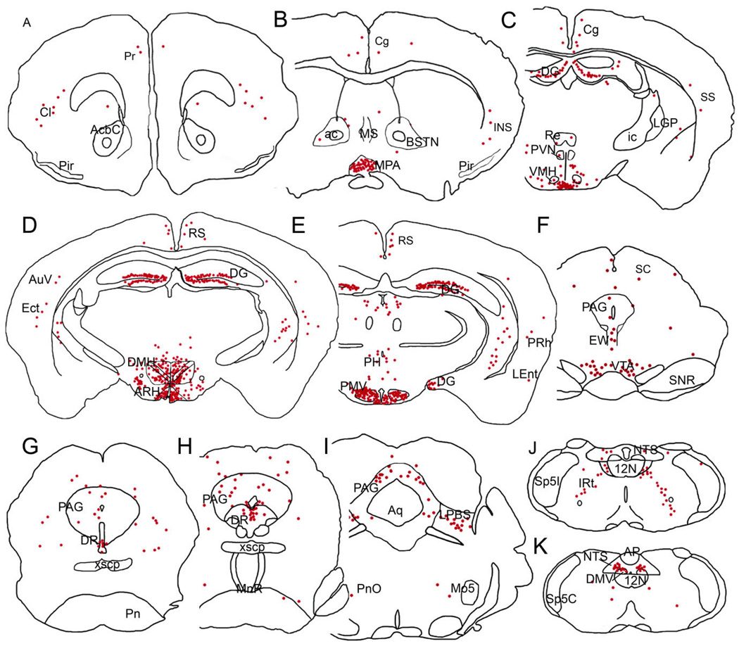
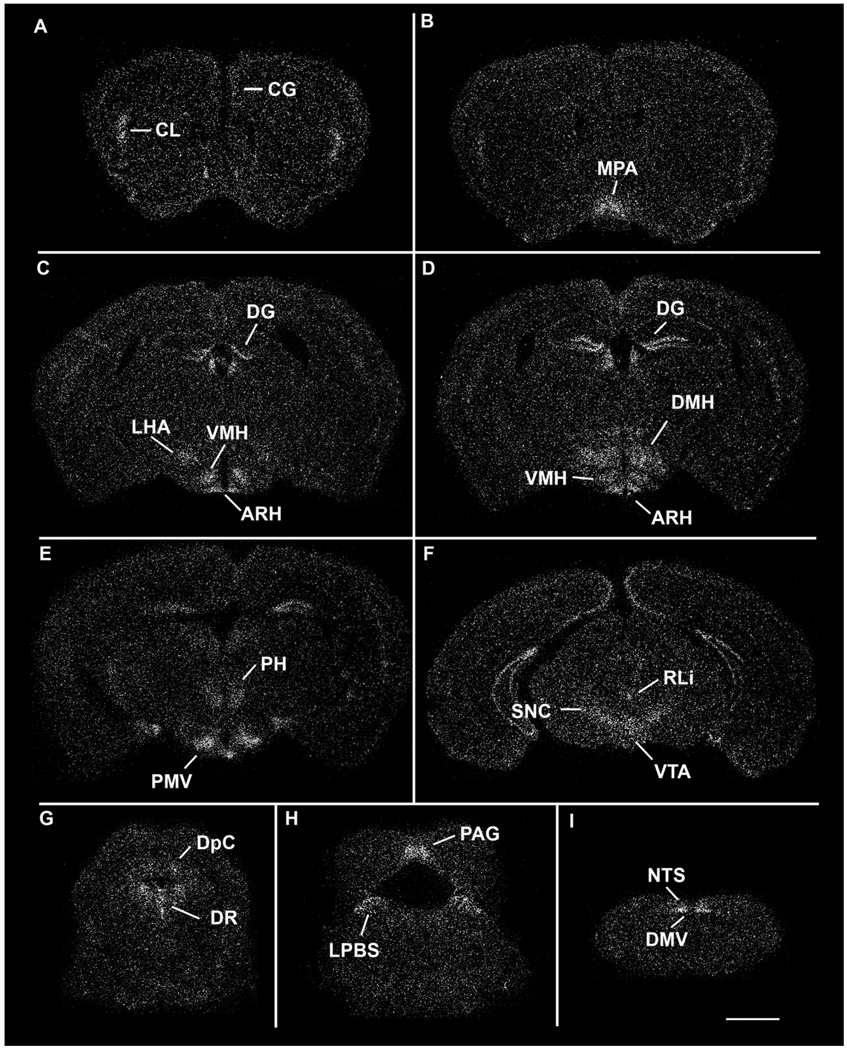
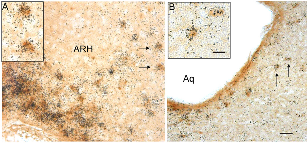
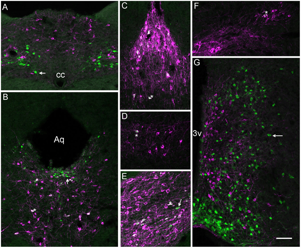
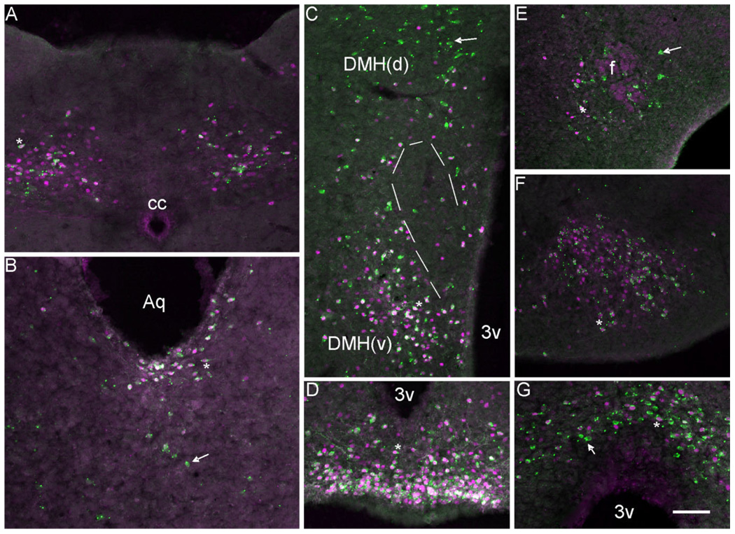
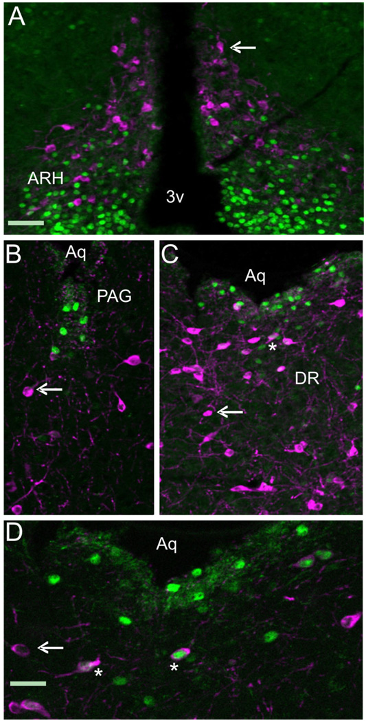
The central actions of leptin are essential for homeostatic control of adipose tissue mass, glucose metabolism, and many autonomic and neuroendocrine systems. In the brain, leptin acts on numerous different cell types via the long-form leptin receptor (LepRb) to elicit its effects. The precise identification of leptin's cellular targets is fundamental to understanding the mechanism of its pleiotropic central actions. We have systematically characterized LepRb distribution in the mouse brain using in situ hybridization in wildtype mice as well as by EYFP immunoreactivity in a novel LepRb-IRES-Cre EYFP reporter mouse line showing high levels of LepRb mRNA/EYFP coexpression. We found substantial LepRb mRNA and EYFP expression in hypothalamic and extrahypothalamic sites described before, including the dorsomedial nucleus of the hypothalamus, ventral premammillary nucleus, ventral tegmental area, parabrachial nucleus, and the dorsal vagal complex. Expression in insular cortex, lateral septal nucleus, medial preoptic area, rostral linear nucleus, and in the Edinger-Westphal nucleus was also observed and had been previously unreported. The LepRb-IRES-Cre reporter line was used to chemically characterize a population of leptin receptor-expressing neurons in the midbrain. Tyrosine hydroxylase and Cre reporter were found to be coexpressed in the ventral tegmental area and in other midbrain dopaminergic neurons. Lastly, the LepRb-IRES-Cre reporter line was used to map the extent of peripheral leptin sensing by central nervous system (CNS) LepRb neurons. Thus, we provide data supporting the use of the LepRb-IRES-Cre line for the assessment of the anatomic and functional characteristics of neurons expressing leptin receptor.


Figures

Figure 1
Figure 1
Generation of mice expressing EYFP/LacZ selectively in LepRb-expressing cells. A: Schematic illustration of the LepRb-IRES-Cre construct. An IRES-NLS-Cre cassette was inserted just 3′ to the LepRb stop codon in the 3′ untranslated region of the LepR gene allowing Cre recombinase to be expressed from the IRES element only in LepRb-containing cells. B: The design for the R26R-EYFP/LacZ Cre recombinase reporter mouse. A floxed transcriptional termination sequence (tp-A) followed by the EYFP or lacZ gene was targeted to the ROSA26 locus. In the absence of Cre recombinase, the tp-A prevents EYFP or lacZ from being expressed. After crossing these mice with a Cre recombinase-expressing mouse strain, a Cre recombination event excises the tp-A sequence, thereby allowing EYFP or LacZ to be expressed under the constitutively active ROSA26 locus selectively in the Cre-producing cell types.
Figure 2
Figure 2
A series of line drawings illustrating the GFP immunoreactivity distribution in the LepRb-EYFP mouse brain. Sections are arranged in a rostral-to-caudal manner (A–K). Each red dot represents ≈2 GFP-IR cells. 12N, hypoglossal nucleus; ac, anterior commissure; AcbC, accumbens nucleus, core; AP, area postrema; Aq, aqueduct; ARH, arcuate nucleus; AuV, secondary auditory nucleus; BSTN, bed nucleus of the stria terminalis; Cg, cingulated cortex; Cl, claustrum; DG, dentate gyrus; DMH, dorsomedial hypothalamic nucleus; DMV, dorsal motor nucleus of the vagus; DR, dorsal raphe; Ect, ectorhinal cortex; EW, Edinger-Westphal nucleus; ic, internal capsule; IRt, intermediate reticular nucleus; LEnt, lateral entorhinal cortex; LHA, lateral hypothalamic area; LGP, lateral globus pallidus; LPBS, superior lateral parabrachial subnucleus; MnR, median raphe nucleus; Mo5, motor trigeminal nucleus; MPA, medial preoptic area; MS, medial septal nucleus; NTS, nucleus of the solitary tract; PAG, periaqueductal gray; PH, posterior hypothalamic area; Pir, piriform cortex; PMV, ventral premammillary nucleus; Pn, pontine nuclei; PnO, pontine reticular nucleus, oral part; PVN, paraventricular hypothalamic nucleus; Re, reuniens thalamic nucleus; RS, retrosplenial cortex; SC, superior colliculus; SNR, substantia nigra, reticular part; Sp5C, spinal trigeminal nucleus, caudal part; Sp5I, spinal trigeminal nucleus, interpolar part; SS, somatosensorial cortex; VMH, ventromedial hypothalamic nucleus; VTA, ventral tegmental area; xscp, decussation of the superior cerebellar peduncle.
Figure 3
Figure 3
A series of low-power darkfield autoradiographs illustrating the distribution of LepRb in the wildtype mouse brain. Sections are arranged in a rostral-to-caudal manner. ARH, arcuate nucleus; Cg, cingulate cortex; Cl, claustrum; DG, dentate gyrus; DMH, dorsomedial hypothalamic nucleus; DMV, dorsal motor nucleus of the vagus; DpC, deep gray layer of the superior colliculus; LPBS, superior lateral parabrachial subnucleus; NTS, nucleus of the solitary tract; PAG, periaqueductal gray; PH, posterior hypothalamic area; PMV, ventral premammillary nucleus; RLi, rostral linear nucleus; SNC, substantia nigra, compact part; VMH, ventromedial hypothalamic nucleus; VTA, ventral tegmental area. Scale bar = 1 mm.
Figure 4
Figure 4
Photomicrographs illustrating the colocalization of LacZ-immunoreactivity and LepRb mRNA in the hypothalamus and midbrain of the LepRb-Cre LacZ mouse. A: Cells in the arcuate nucleus of the hypothalamus (ARH). B: Cells in the dorsal raphe of LepRb-Cre LacZ tissue coexpressing LepRb mRNA and LacZ. Cells labeled with arrows denote examples of double-labeled cells presented at higher magnification in the panel inserts. Cells containing LepRb mRNA were labeled with silver grain clusters and LacZ-IR cells are labeled with a brown cytoplasmic precipitate. Scale bar: = 50 µm in A,B; 25 µm in inserts.
Figure 5
Figure 5
A series of photomicrographs illustrating the coexpression of tyrosine hydroxylase (TH) and LepRb. A–G: LepRb-Cre LacZ sections labeled for TH (magenta) and LacZ (green) in (A) nucleus of the solitary tract (NTS), (B) the dorsal raphe nucleus (DR) and periaqueductal gray (PAG), (C) in the caudal linear nucleus (Cli), (D) in the A8 nucleus, (E) in the ventral tegmental area (VTA), (F) in the substantia nigra, compact part (SNC), and (G) in the mediobasal hypothalamus. Single-labeled cells appear green or magenta (arrow) while coexpression (asterisk) is indicated by cells appearing white due to color overlay or magenta/white with green nuclei. Aq, aqueduct; 3v, third ventricle; cc, central canal. Scale bar =100 µm.
Figure 6
Figure 6
A series of photomicrographs illustrating the coexpression of LepRb-Cre LacZ with pSTAT3. A–G: LepRb-Cre LacZ sections labeled for pSTAT3 (magenta) and LacZ (green). Coexpression was observed in (A) nucleus of the solitary tract (NTS), (B) in the dorsal raphe (DR) and periaqueductal gray (PAG), (C) in the DMH(d): dorsal medial hypothalamus dorsal part and DMH(v) dorsal medial hypothalamus ventral part, (D) in the arcuate nucleus (ARH), (E) in the lateral hypothalamus, (F) in the ventral premammilary nucleus (PMV), and (G) in the medial preoptic nuclei (MPO). Single-labeled cells appear green with uniform or punctuate cytoplasmic expression. Double-labeled cells have magenta/white nuclei with punctuate or uniform green cytoplasm. PMV double-labeled neurons are almost exclusively punctuate with respect to LacZ expression. Aq, aqueduct; 3v, third ventricle; cc, central canal; f, fornix. Scale bar =100 µm.
Figure 7
Figure 7
A series of photomicrographs illustrating the coexpression of TH with pSTAT3. A–D: LepRb-Cre LacZ sections labeled for TH (magenta) and pSTAT3 (green) in (A) arcuate nucleus (ARH), (B) periaqueductal gray (PAG), (C,D) dorsal raphe (DR). Single-labeled cells are magenta or green (arrow), while double-labeled cells have a green/white nucleus surrounded by magenta cytoplasm (asterisk). 3v: third ventricle, Aq: aqueduct. Scale bars = 100 µm for A–C; 50 µm in D.

References

    1. Ahima RS, Saper CB, Flier JS, Elmquist JK. Leptin regulation of neuroendocrine systems. Front Neuroendocrinol. 2000;21:263–307. - PubMed
    1. Badman MK, Flier JS. The gut and energy balance: visceral allies in the obesity wars. Science. 2005;307:1909–1914. - PubMed
    1. Balthasar N, Coppari R, McMinn J, Liu SM, Lee CE, Tang V, Kenny CD, McGovern RA, Chua SC, Jr, Elmquist JK, Lowell BB. Leptin receptor signaling in POMC neurons is required for normal body weight homeostasis. Neuron. 2004;42:983–991. - PubMed
    1. Bates SH, Stearns WH, Dundon TA, Schubert M, Tso AW, Wang Y, Banks AS, Lavery HJ, Haq AK, Maratos-Flier E, Neel BG, Schwartz MW, Myers MG., Jr STAT3 signalling is required for leptin regulation of energy balance but not reproduction. Nature. 2003;421:856–859. - PubMed
    1. Benoit SC, Clegg DJ, Seeley RJ, Woods SC. Insulin and leptin as adiposity signals. Recent Prog Horm Res. 2004;59:267–285. - PubMed

Publication types