. 2009 Apr;27(4):783-95.
doi: 10.1002/stem.13.

Derivation of primordial germ cells from human embryonic and induced pluripotent stem cells is significantly improved by coculture with human fetal gonadal cells

Affiliations

Derivation of primordial germ cells from human embryonic and induced pluripotent stem cells is significantly improved by coculture with human fetal gonadal cells

Tae Sub Park et al. Stem Cells. 2009 Apr.

Abstract

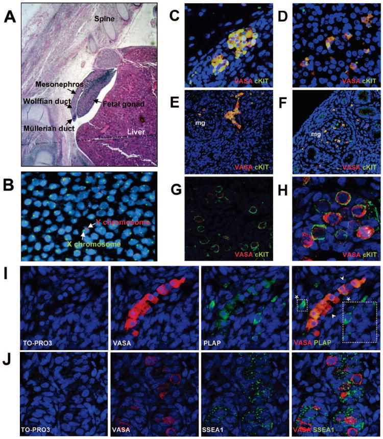
The derivation of germ cells from human embryonic stem cells (hESCs) or human induced pluripotent stem (hIPS) cells represents a desirable experimental model and potential strategy for treating infertility. In the current study, we developed a triple biomarker assay for identifying and isolating human primordial germ cells (PGCs) by first evaluating human PGC formation during the first trimester in vivo. Next, we applied this technology to characterizing in vitro derived PGCs (iPGCs) from pluripotent cells. Our results show that codifferentiation of hESCs on human fetal gonadal stromal cells significantly improves the efficiency of generating iPGCs. Furthermore, the efficiency was comparable between various pluripotent cell lines regardless of origin from the inner cell mass of human blastocysts (hESCs), or reprogramming of human skin fibroblasts (hIPS). To better characterize the iPGCs, we performed Real-time polymerase chain reaction, microarray, and bisulfite sequencing. Our results show that iPGCs at day 7 of differentiation are transcriptionally distinct from the somatic cells, expressing genes associated with pluripotency and germ cell development while repressing genes associated with somatic differentiation (specifically multiple HOX genes). Using bisulfite sequencing, we show that iPGCs initiate imprint erasure from differentially methylated imprinted regions by day 7 of differentiation. However, iPGCs derived from hIPS cells do not initiate imprint erasure as efficiently. In conclusion, our results indicate that triple positive iPGCs derived from pluripotent cells differentiated on hFGS cells correspond to committed first trimester germ cells (before 9 weeks) that have initiated the process of imprint erasure.


Figures

Figure 1
Figure 1. Surface markers that define male human primordial germ cell (PGC) migration and colonization in the first trimester of human fetal life
(A) Hematoxylin and Eosin staining of a 5 week human embryo. (B) Fluorescent in situ hybridization (FISH) with X chromosome-specific probe (green) and Y chromosome-specific probe (red) to determine embryo sex. (C-H) Immunofluorescent staining of overlay images showing the germ cell-specific marker VASA (red) and cKIT (green). Co-localization is seen as yellow. (C) VASA/cKIT co-expression (yellow) in PGC clusters exiting the hind gut (800x) (D) VASA/cKIT co-expression in PGCs migrating towards gonad through the mesonephros which contains mesonephric glomeruli (mg) (800x) (E) VASA/cKIT double positive PGCs entering into the gonad as a cluster from the mesonephros (400x) (F) VASA/cKIT double positive PGCs migrating through the mesonephric glomeruli (mg) during migration to the gonad (400x) (G,H) VASA/cKIT double positive PGCs in the fetal gonad (800x and 1600x) (I) Migrating VASA (red) positive PGCs with PLAP (green) (800x). (*) indicate PLAP positive VASA negative cells which were only observed during migration. (J) VASA (red) with SSEA1 (green) in colonized PGCs within the fetal gonad (1200x). TO-PRO 3 iodide was used as a marker for cell nucleus in all sections from B-J.
Figure 2
Figure 2. SSEA1 co-localizes with cKIT positive cells in the first trimester fetal gonad
(A) Human fetal testis at 9 weeks of gestation AMH (red) SSEA1 (green), TO-PRO 3 iodide stains the cell nucleus (1200x). Arrow indicates exclusive red and arrowhead indicates exclusive green cells. (B) Flow analysis of a 7-gestational week-old male gonad. Shown are negative controls (red lines) cKIT and SSEA1, (blue lines).
Figure 3
Figure 3. Isolation and characterization of stromal cells from first trimester human fetal gonads and comparison of different feeder layers for iPGC differentiation
(A) hFGS cells grow as a monolayer and are negative for Alkaline Phosphatase (AP), whereas hESCs are positive for AP. (B) RT-PCR comparing hFGS cells and adult testis. (C) Flow cytometry of hFGS cells for TRA-1-85 (pan-human surface marker), SSEA1, cKIT and PLAP. (D) Percentages of cKIT/SSEA1/VASA triple positive cells in undifferentiated (Undiff.) HSF-1 and HSF-6 hESCs, and following differentiation for three days on matrigel, hFGS cells or matrigel with hFGS cell-conditioned media. (E) Comparison of iPGC differentiation in triplicate on hFGS cells, human placental stromal cells and human liver stromal cells after 7-day differentiation. Significance was achieved when comparing a to b and c to d (P<0.05).
Figure 4
Figure 4. Derivation of iPGCs from HSF-1, HSF-6 and H9 lines of hESCs using hFGS cells consistently results in formation of triple positive cells at day 7 of differentiation
(A) Flow cytometry of the HSF-1 line of hESCs. Red lines indicate negative controls (secondary antibody only) and blue lines indicate positive staining for each molecule as indicated above each plot. Following 7 days of differentiation, cKIT, SSEA1, PLAP and VASA positive cells could be identified above the negative control. Analysis of two channels reveals presence of double positive cKIT/SSEA1 and PLAP/SSEA1 cells. VASA expression was re-analyzed in the double positive (blue) and negative (red) populations after 7 days of differentiation. This demonstrates that VASA positive cells are only found within the cKIT/SSEA1 or PLAP/SSEA1 double positive population and not the netative. (B) Quantification of iPGC formation from HSF-1, HSF-6 and H9 lines of hESC lines at day 7 of differentiation. All experiments were performed in triplicate. Significance was achieved when comparing a to b and c to d (P<0.05). (C) Relative expression of VASA to GAPDH mRNA using Real-time PCR in double-positive (cKIT/SSEA1) cells sorted by FACS at day 7 of differentiation from HSF1 and HSF6 lines of hESCs (left panel). Middle panel shows a gel electrophoresis of the PCR products of VASA RT-PCR in the cKIT/SSEA1 double positive and double negative FACS sorted cells at day 7 of differentiation from HSF1 (middle). Immunofluorescent staining of HSF1-derived PGCs with cKIT (green) and VASA (red) after MACS-sorting and cytospin. VASA protein is expressed in the cytoplasm of cKIT-positive PGCs (1200x) (right panel).
Figure 5
Figure 5. Gene expression and CpG methylation status at imprinted genes in isolated iPGCs at day 7 of differentiation from HSF-1 and HSF-6
(A) Heat map showing differentially expressed genes in HSF-1 and HSF-6 cKIT/SSEA1 double positive FACS sorted cells compared to the double negative cells. Real time PCR of germ cell-related genes (PRDM1, DPPA3, DAZL) and somatic genes (HOXA2, HOXC5) in double-positive (cKIT/SSEA1) and double-negative FACS sorted cells from HSF-1 at day 7 of differentiation. (B) CpGs of imprinted genes (H19, PEG1 and SNRPN DMRs) were analyzed following bisulfite sequencing from undifferentiated HSF-1 and HSF-6 hESCs and iPGCs derived at day 7 of differentiation. In vitro derived PGCs were isolated by FACS sorting for cKIT/SSEA1 double positive cells. Methylated CpGs are represented as filled circles, unmethylated CpGs are represented as open circles. Percentage methylation was calculated comparing the number of CpG methylated sites in undifferentiated hESCs before differentiation with the number of CpG methylated sites at the same locus in the iPGC population. Decreased methylation in iPGCs relative to undifferentiated hESCs is shown in green and no change shown in black. Abnormal methylation is shown in red.
Figure 6
Figure 6. Derivation of iPGCs from hIPS cells and status of CpG methylation at imprinted genes in the hIPS-derived iPGCs
(A) Quantification of iPGC derived from two hIPS lines (hIPS1, hIPS2) following flow analysis. Significance was achieved when comparing a to b and c to d (P<0.05). (C) CpGs of imprinted genes (H19, PEG1 and SNRPN DMRs) were analyzed by bisulfite sequencing from fibroblasts before reprogramming, undifferentiated hIPS2 and iPGCs sorted at day 7 of differentiation using cKIT/SSEA1 surface markers. The cKIT/SSEA1 double negative population was used as a control. Methylated CpGs are represented as filled circles, unmethylated CpGs are represented as open circles. Percentage methylation was calculated by comparing the number of CpG methyl groups in the starting population verses the test group. For example fibroblast verses hIPS2; hIPS2 verses cKIT/SSEA1 FACS sorted double positive iPGC; and hIPS2 verses the double negative population. Decreased methylation is shown in green no change shown in black and abnormal methylation is shown in red.

References

    1. Daley GQ. Gametes from embryonic stem cells: a cup half empty or half full? Science. 2007 Apr 20;316(5823):409–410. - PubMed
    1. Holden C. Sperm from Skin Becoming a Reality. ScienceNOW daily News. 2008;415:2.
    1. Surani MA. Germ cells: the eternal link between generations. C R Biol. 2007 Jun-Jul;330(6-7):474–478. - PubMed
    1. Hayashi K, De Sousa Lopes S, Surani M. Germ cell specification in mice. Science. 2007;316:394–396. - PubMed
    1. Ying Y, L XM, Marble A, Lawson KA, Zhao GQ. Requirement of Bmp8b for the generation of primordial germ cells in the mouse. Mol Endocrinol. 2000;14:1053–1063. - PubMed

Publication types

MeSH terms