. 2009 Apr;27(4):866-77.
doi: 10.1002/stem.2.

Stem cell property of postmigratory cranial neural crest cells and their utility in alveolar bone regeneration and tooth development

Affiliations

Stem cell property of postmigratory cranial neural crest cells and their utility in alveolar bone regeneration and tooth development

Il-Hyuk Chung et al. Stem Cells. 2009 Apr.

Abstract

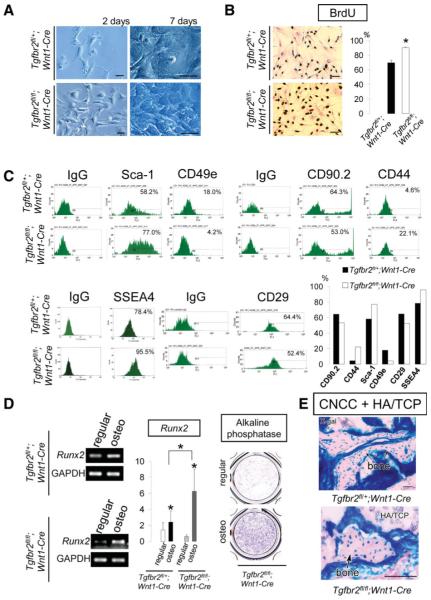
The vertebrate neural crest is a multipotent cell population that gives rise to a variety of different cell types. We have discovered that postmigratory cranial neural crest cells (CNCCs) maintain mesenchymal stem cell characteristics and show potential utility for the regeneration of craniofacial structures. We are able to induce the osteogenic differentiation of postmigratory CNCCs, and this differentiation is regulated by bone morphogenetic protein (BMP) and transforming growth factor-beta signaling pathways. After transplantation into a host animal, postmigratory CNCCs form bone matrix. CNCC-formed bones are distinct from bones regenerated by bone marrow mesenchymal stem cells. In addition, CNCCs support tooth germ survival via BMP signaling in our CNCC-tooth germ cotransplantation system. Thus, we conclude that postmigratory CNCCs preserve stem cell features, contribute to craniofacial bone formation, and play a fundamental role in supporting tooth organ development. These findings reveal a novel function for postmigratory CNCCs in organ development, and demonstrate the utility of these CNCCs in regenerating craniofacial structures.


Figures

Figure 1
Figure 1
Stem cell characteristics of postmigratory CNCCs. (A): Postmigratory CNCCs cultured at a low cell density (5 × 10³ cells in Ø 10-cm culture dish) for 3 weeks formed adherent clonogenic cell clusters (arrow head), visualized with X-gal staining. Top: The culture dish with several X-gal+ colonies. Arrowhead indicates a colony. Bottom: Magnified image of an X-gal+ colony. (B): Immunostaining of colonies with α-SMA, Type I collagen, NF145, and S100. (C): BrdU incorporation assay of multicolony-derived CNCCs and BMMSCs. Right: Quantitation of BrdU incorporation in CNCCs and BMMSCs (*, p < .05). (D): Fluorescence-activated cell sorting (FACS) analysis of P1 postmigratory CNCC and BMMSC cultures. Right: Quantitation of FACS analysis. Scale bars = 100 μm (A, B), 50 μm (C). Abbreviations: BMMSCs, bone marrow mesenchymal stem cells; BrdU, 5-bromo-2prime-deoxyurine; CNCCs, cranial neural crest cells; FDG, fluorescein di-β-d-galactopyranoside; NF145, neurofilament 145; PE, phycoerythrin; Sca-1, stem cell antigen-1; α-SMA, α-smooth muscle actin; SSEA4, stage-specific embryonic antigen 4; T Blue, toluidine blue.
Figure 2
Figure 2
Multipotent differentiation of postmigratory CNCCs. (A, B): Osteogenic differentiation of postmigratory CNCCs and BMMSCs. Alkaline phosphatase (A) and alizarin red (B) analysis of CNCCs and BMMSCs following culture in regular or osteogenic induction medium. Quantitation indicates elevated expression of alkaline phosphatase (A) and calcified nodule (B) formation in CNCCs following osteogenic induction (***, p < .001), and increased calcium concentration (B, * , p < .05). (C, D): Adipogenic differentiation of CNCCs and BMMSCs. Oil Red O staining following culture in regular or adipogenic induction medium (C). Reverse transcription polymerase chain reaction of adipogenic markers PPARγ2 and LPL of CNCCs following 2-weeks culture in regular or adipogenic induction medium ([D], top). Quantitation of lipid droplets in CNCCs and BMMSCs ([D], bottom). (E): H&E staining (a, b), toluidine blue staining (c, d), and type II collagen immunostaining (e, f) of CNCCs and BMMSCs following 4 weeks chondrogenic differentiation. (F): Analysis 8 weeks after subcutaneous transplantation of CNCCs and HA/TCP into immunocompromised mice. Bone matrix was detected with H&E staining (a). The osteocytes located in the bone matrix were positive for β-galactosidase activity after X-gal staining (b, c, e). Alizarin red (d) and von Kossa (g) staining and expression of alkaline phosphatase (h) and osteocalcin (i) indicate bone formation. Black arrow heads (b, c, e, f) indicate CNCCs-derived cells. (E): H&E (a, b), X-gal (c, d), Masson’s trichrome (e, f), and picrosirius red (g, h) analysis 8 weeks after transplantation of CNCCs with Matrigel or Gelfoam. The transplants of CNCC and Matrigel resulted in tendon-like structures [(a), arrowheads, dashed line circles] with dense collagen matrix deposition ([e, g] arrows). The CNCCs transplanted with Gelfoam also resulted in condensed collagen matrix ([b, f, h], arrows, dashed line). Scale bars = 50 μm. Abbreviations: BMMSCs, bone marrow mesenchymal stem cells; B.V., blood vessel; CNCCs, cranial neural crest cells; C.T., connective tissue; HA/TCP, hydroxyapatite/tricalcium phosphate; LPL, lipoprotein lipase; PPARγ2, peroxisome proliferator-activated receptor gamma 2.
Figure 3
Figure 3
Self-renewal of postmigratory CNCCs. (A–D): Transplantderived CNCC cells retain stem cell characteristics. (A): Strategy for evaluating self-renewal of postmigratory CNCCs [21]. (B): After in vitro and in vivo expansion CNCCs can form colonies (arrow head) when cultured at a low cell density. (C): Alizarin red and Oil Red O staining of transplant-derived CNCCs. (D): X-gal staining of transplant-derived CNCCs (arrow heads) retransplanted with HA/TCP indicate they retain their ability to form bone in vivo. (E–H): The contribution of CNCC-originated cells to the MSCs of the mandible of Wnt1-Cre;R26R mice. (E): CNCC-originated cells can form clonogenic cell clusters (arrowhead), visualized with X-gal staining. (F): Fluorescence-activated cell sorting analysis of CNCC-originated mandibular MSCs. (G): Osteogenic and adipogenic induction of CNCC-originated MSCs, stained with Alizarin red and Oil Red O, respectively. (H): CNCC-originated MSCs (arrow heads on X-gal staining) can form bone when subcutaneously transplanted with HA/TCP. Scale bars = 100 μm (B, C, E), 50 μm (D, G, H). Abbreviations: BA, branchial arch; CNCCs, cranial neural crest cells; FDG, fluorescein di-β-d-galactopyranoside; HA/TCP, hydroxyapatite/tricalcium phosphate; MSCs, mesenchymal stem cell; S.C., subcutaneous transplantation; Sca-1, stem cell antigen-1; SSEA4, stage-specific embryonic antigen 4.
Figure 4
Figure 4
CNCCs have distinct bone-forming properties. (A, B): Histological analysis of a defect in the calvaria in mice 8 weeks after transplantation of no graft (b), CNCCs and HA/TCP (c, d), or BMMSCs and HA/TCP (e, f). Arrowheads indicate the margin of the calvarial defect. (a) Graft into the calvarial defect. (b) No bone formation is detectable by H&E staining in mice that received no graft. (c, d) After transplantation of CNCCs and HA/TCP, bone matrix can be detected by H&E staining (arrows) that is similar to the host predefect calvaria (c). The newly formed bone matrix contained X-gal positive osteocytes (d). (e, f) Bone can be visualized by H&E staining (arrow) after BMMSCs and HA/TCP transplantation that contains prominent bone marrow space development (f). (B): Quantitative analysis indicated that the formation of mineralized matrix was greater in CNCC transplants (*, p < .05), but the formation of bone marrow was greater in BMMSC transplants (*, p < .05). (C): Quantitative reverse transcription polymerase chain reaction of Runx2 and Osteopontin expression in CNCCs cultured in regular medium, osteogenic induction medium or osteogenic induction medium, and exogenous BMP2. Osteogenic induction medium increased the expression of Runx2 and Osteopontin, and the addition of BMP2 enhanced these effects (*, p < .05). (D): Immunostaining of Osteocalcin in CNCCs cultured in regular or osteogenic induction medium, alone or with the addition of BMP2, TGF-β2, or FGF9. (E): H&E and X-gal staining and CD45 immunohistochemistry of CNCCs and HA/TCP transplanted with BMP2 beads. Bone marrow space is indicated with arrow heads. (B): BMP2. (F): H&E staining of transplants with TGF-β2 beads. Note fibrous tissues (arrow heads) mainly composed of collagen fibers and the absence of bone matrix formation. (G): H&E staining of transplants with FGF9 beads. Note small amount of bone matrix and abundant blood vessel formation. (F): FGF9 beads. Scale bars = 100 μm (D), 50 μm (A, E–G). Abbreviations: BMMSCs, bone marrow mesenchymal stem cells; BMP2, bone morphogenetic protein 2; CNCCs, cranial neural crest cells; C.T., connective tissue; FGF9, fibroblast growth factor 9; HA/TCP, hydroxyapatite/tricalcium phosphate; TGF-β2, transforming growth factor-β2.
Figure 5
Figure 5
Transforming growth factor-β (TGF-β) signaling regulation of postmigratory CNCCs. (A): Tgfbr2fl/+;Wnt1-Cre (control) and Tgfbr2fl/fl;Wnt1-Cre CNCCs after 2 and 7 days of culture. (B): BrdU incorporation analysis of Tgfbr2fl/+;Wnt1-Cre (control) and Tgfbr2fl/fl;Wnt1-Cre CNCCs. Statistical analysis indicates that proliferation was higher in CNCCs lacking TGF-β (***, p < .001). (C): Fluorescence-activated cell sorting analysis of Tgfbr2fl/+;Wnt1-Cre (control) and Tgfbr2fl/fl;Wnt1-Cre CNCCs. (D): Analysis of osteogenic marker gene expression. Expression of Runx2, by reverse transcription polymerase chain reaction, and alkaline phosphatase was assayed in regular and osteogenic induction (osteo) media. Statistical analysis indicates that Runx2 is elevated in CNCCs lacking TGF-β type II receptors (*, p < .05). (E): Bone matrix formation (arrows) was detected by X-gal staining in Tgfbr2fl/+;Wnt1-Cre (control) and Tgfbr2fl/fl;Wnt1-Cre CNCCs + HA/TCP transplants. Scale bars = 50 μm. Abbreviations: BrdU, 5-bromo-2′-deoxyurine; BMMSCs, bone marrow mesenchymal stem cells; CNCCs, cranial neural crest cells; HA/TCP, hydroxyapatite/tricalcium phosphate; Sca-1, stem cell antigen-1; SSEA4; stage-specific embryonic antigen 4.
Figure 6
Figure 6
Postmigratory CNCCs support tooth germ survival. Analysis of tooth germs transplanted with HA/TCP alone (A, B) or with CNCCs (C–G), or BMMSCs (H, I). (A, B): H&E staining indicates that tooth germs transplanted with HA/TCP alone gave rise to keratinized cysts ([A], arrowheads) and intramembranous bone formation ([B], arrows). (C–G): H&E (C) and X-gal staining (D) of tooth germs transplanted with CNCCs and HA/TCP reveal apparently normal teeth. Transplanted CNCCs were detectable within the dental pulp ([D], Lower inset: magnified image, arrow heads) and alveolar bone ([F], arrow heads) after X-gal staining. TRAP-positive staining was present in the bone marrow space and blood vessels ([G], arrows). (H, I): Bone matrix formation can be detected by X-gal staining in tooth germs from Wnt1-Cre;R26R mice transplanted with BMMSCs and HA/TCP (H). The osteocytes in the bone matrix are positive for β-galactosidase activity after X-gal staining ([H], arrowheads). Two tooth germs out of twelve transplanted resulted in abnormal tooth formation (I). Scale bars = 50 μm. Abbreviations: Alv. Bone, alveolar bone; BMMSCs, bone marrow mesenchymal stem cells; CNCCs, cranial neural crest cells; D, dentin; E, enamel; HA/TCP, hydroxyapatite/tricalcium phosphate; P, pulp; PDL, periodontal ligament.
Figure 7
Figure 7
BMP signals play a role in CNCCs-mediated tooth development. (A–C): H&E staining of T.G. transplanted with HA/TCP and BMP4 beads (A), BMP4 and ActivinβA beads (B), or BMP4, ActivinβA, FGF2, and FGF4 beads (C). The T.G. gave rise to cysts and bone. Arrowheads indicate bone. (D): X-gal staining of T.G. transplanted with CNCCs, HA/TCP, and BMP4, or Noggin beads. (E): X-gal staining of tooth germs transplanted with CNCCs from Smad4fl/fl;Wnt1-Cre mice and HA/TCP. Transplanted tooth germs gave rise to intramembranous bone formation and keratinized cysts. (F): Semiquantitative reverse transcription polymerase chain reaction analysis of control or Smad4-siRNA-treated CNCCs (left). X-gal and H&E staining of tooth germs transplanted with Smad4-siRNA-treated CNCCs and HA/TCP. Note that the treatment of CNCCS with Smad4-siRNA resulted in abnormal teeth or cysts. (G): H&E staining of T.G. transplanted with BMMSCs, BMP4 beads, and HA/TCP. These T.G. resulted in cyst and bone matrix formation. (B): BMP4 beads. Scale bars = 50 μm. Abbreviations: Alv. Bone, Alveolar bone; B, BMP4 beads; BMMSCs, bone marrow mesenchymal stem cells; BMP4, bone morphogenetic protein 4; CNCCs, cranial neural crest cells; FGF2, fibroblast growth factor 2; FGF4, fibroblast growth factor 4; GF, growth factor containing beads; HA/TCP, hydroxyapatite/tricalcium phosphate; N, noggin beads; PDL, periodontal ligament.

References

    1. Noden DM. Cell movements and control of patterned tissue assembly during craniofacial development. J Craniofac Genet Dev Biol. 1991;11:192–213. - PubMed
    1. Le Douarin NM, Ziller C, Coul G. Patterning of neural crest derivatives in the avian embryo: In vivo and in vitro studies. Dev Biol. 1993;159:24–49. - PubMed
    1. Trainor PA, Krumlauf R. Patterning the cranial neural crest: hindbrain segmentation and Hox gene plasticity. Nat Rev Nurosci. 2000;1:116–124. - PubMed
    1. Chai Y, Jiang X, Ito Y, et al. Fate of the mammalian cranial neural crest during tooth and mandibular morphogenesis. Development. 2000;127:1671–1679. - PubMed
    1. Chai Y, Maxson RE., Jr Recent advances in craniofacial morphogenesis. Dev Dyn. 2006;235:2353–2375. - PubMed

Publication types

MeSH terms

Substances