. 2009 Mar 1;43(5):1605-11.
doi: 10.1021/es801925c.

Quantum dot nanotoxicity assessment using the zebrafish embryo

Affiliations

Quantum dot nanotoxicity assessment using the zebrafish embryo

Tisha C King-Heiden et al. Environ Sci Technol. .

Abstract

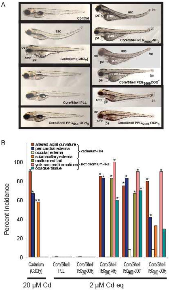
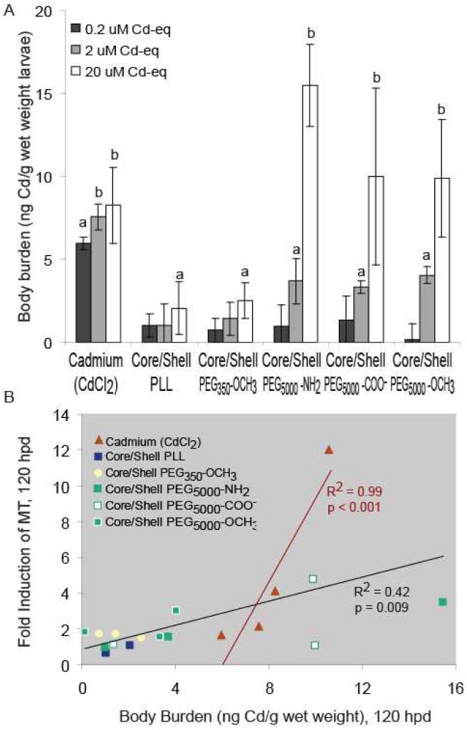
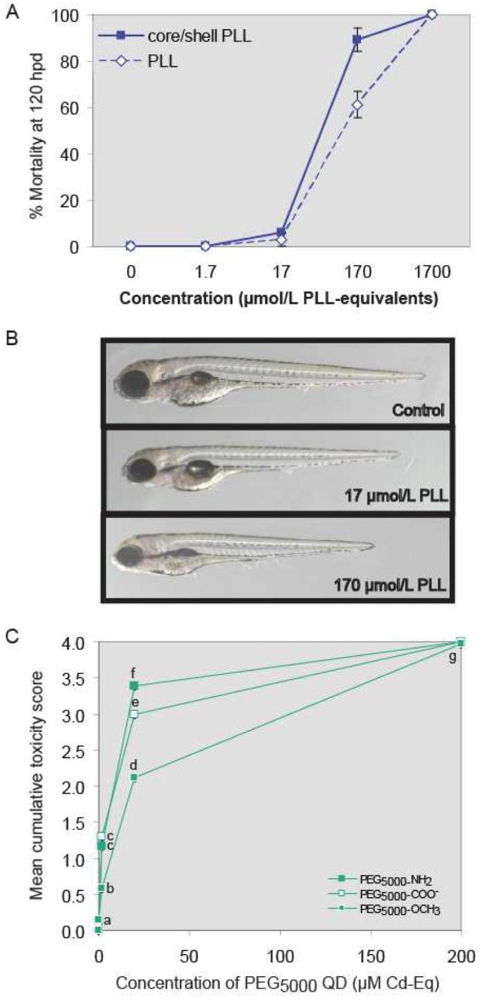
Quantum dots (QDs) hold promise for several biomedical, life sciences, and photovoltaic applications. Substantial production volumes and environmental release are anticipated. QD toxicity may be intrinsic to their physicochemical properties, or result from the release of toxic components during breakdown. We hypothesized that developing zebrafish could be used to identify and distinguish these different types of toxicity. Embryos were exposed to aqueous suspensions of CdSe(core)/ZnS(shell) QDs functionalized with either poly-L-lysine or poly(ethylene glycol) terminated with methoxy, carboxylate, or amine groups. Toxicity was influenced by the QD coating, which also contributed to the QD suspension stability. At sublethal concentrations, many QD preparations produced characteristic signs of Cd toxicity that weakly correlated with metallothionein expression, indicating that QDs are only slightly degraded in vivo. QDs also produced distinctly different toxicity that could not be explained by Cd release. Using the zebrafish model, we were able to distinguish toxicity intrinsic to QDs from that caused by released metal ions. We conclude that developing zebrafish provide a rapid, low-cost approach for assessing structure-toxicity relationships of nanoparticles.


Figures

Figure 1
Figure 1
A. Exposure of zebrafish larvae for 120 h to 20 μM CdCl2 or 2 μM Cd equivalents of different functionalized QDs caused similar mortality (3-6%) but different endpoints of toxicity. CdCl2 caused four toxic responses: altered axial curvature, pericardial edema, ocular edema, and submaxillary edema, whereas functionalized QDs caused a more complex pattern of “cadmium-like” and “not cadmium-like” responses. Endpoints of toxicity that were “not cadmium-like” consisted of: malformed tail, yolk sac malformation (yolk not absorbed), and opaque tissue indicative of tissue necrosis in the head, body, and yolk sac. This combination of “cadmium-like” and “not cadmium-like” responses to functionalized QD exposure suggested that both Cd and the functionalized QD cause toxicity. Abbreviations: aac = altered axial curvature; sme = submandibular edema; pe = pericardial edema; yse = yolk sac edema; ysm = yolk sac malformation; oe = ocular edema; and tm = tail malformation. B. Effect of equally effective mortality-inducing concentrations of CdCl2 (20 μM) or functionalized QDs (2 μM Cd equivalents) in zebrafish on the incidence, above that in unexposed control (not shown), of different endpoints of toxicity. Results across combined replicates (n = 12) are shown for exposure to 20 μM Cd (as CdCl2) or to 2 μM Cd equivalents of different functionalized QDs. Asterisks denote increased incidence above unexposed control (p < 0.05). QD cores were CdSe and the shell was ZnS.
Figure 2
Figure 2
A. Cadmium accumulation following 120-h waterborne exposure to graded concentrations of functionalized QDs or CdCl2. The mean body burden (ngCd·glarvae-1) of unexposed control Zebrafish larvae was subtracted from that of zebrafish larvae exposed to CdCl2 or QDs to determine their Cd body burden. Values represent mean ± SE, n = 3 pools of 10 larvae each; letters denote significant differences (p < 0.05). Those without letters did not differ from unexposed control zebrafish. Insufficient numbers of embryos survived to 120 hpd to allow measurement of body burden following exposure to 200 μM Cd equivalents of QDs. B. Correlation between Cd body burden and MT fold induction relative mRNA abundance after 120-h exposure. CdCl2 exposures (red) were correlated separately from those for QD exposures. Correlations for individual QDs are presented in Table S1. QD cores were composed of CdSe; shells were ZnS.
Figure 3
Figure 3
A. Comparison of mortality induced by PLL-wrapped CdSecore/ZnSshell QDs with that produced by PLL alone. Expressed as PLL equivalents, LC50 values for CdSecore/ZnSshell-PLL QDs and PLL were 59 (44-75) and 125 (95-154) μM (95% confidence intervals in parentheses). B. Representative photomicrographs of larvae exposed to PLL alone. Beyond reduced growth, no sublethal toxicity was seen. C. Cumulative toxicity scores following 120-h exposure to CdSecore/ZnSshell PEG5000 QDs with different surface chemistries. Values represent mean ± SE (n = 36); letters denote significant differences (p < 0.05). Note: Error bars are present, but smaller than symbols used in the graph.

References

    1. Michalet X, Pinaud FF, Bentolila LA, Tsay JM, Doose S, Li JJ, Sundaresan G, Wu AM, Gambhir SS, Weiss S. Quantum dots for live cells, in vivo imaging, and diagnostics. Science. 2005;307:538–544. - PMC - PubMed
    1. Gratzel M. Photovoltaic and photoelectrochemical conversion of solar energy. Philos. Transact. A Math. Phys. Eng Sci. 2007;365:993–1005. - PubMed
    1. Nirmal M, Brus L. Luminescence photophysics in semiconductor nanocrystals. Acc. Chem. Res. 1999;32:407–414.
    1. Derfus AM, Chan WCW, Bhatia SN. Probing the cytotoxicity of semiconductor quantum dots. Nano Lett. 2004:11–18. - PMC - PubMed
    1. Akerman ME, Chan WC, Laakkonen P, Bhatia SN, Ruoslahti E. Nanocrystal targeting in vivo. Proc. Natl. Acad. Sci U. S. A. 2002;99:12617–12621. - PMC - PubMed

Publication types