

Dorsolateral and dorsomedial prefrontal gray matter density changes associated with bipolar depression
- PMID: 19351579
- PMCID: PMC3265395
- DOI: 10.1016/j.pscychresns.2008.06.007
Dorsolateral and dorsomedial prefrontal gray matter density changes associated with bipolar depression
Abstract
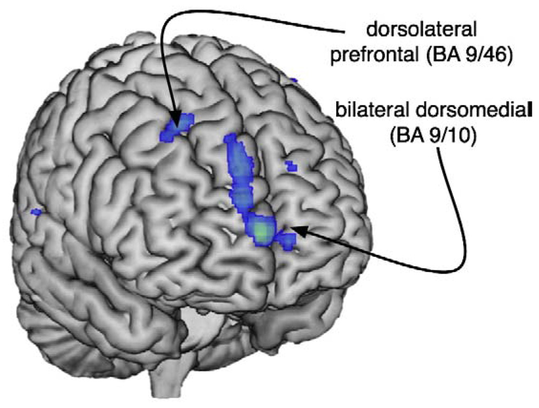
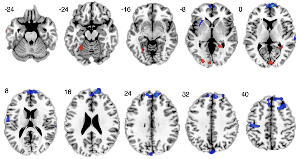
Mood states are associated with alterations in cerebral blood flow and metabolism, yet changes in cerebral structure are typically viewed in the context of enduring traits, genetic predispositions, or the outcome of chronic psychiatric illness. Magnetic resonance imaging (MRI) scans were obtained from two groups of patients with bipolar disorder. In one group, patients met criteria for a current major depressive episode whereas in the other no patient did. No patient in either group met criteria for a current manic, hypomanic, or mixed episode. Groups were matched with respect to age and illness severity. Analyses of gray matter density were performed with Statistical Parametric Mapping software (SPM5). Compared with non-depressed bipolar subjects, depressed bipolar subjects exhibited lower gray matter density in the right dorsolateral and bilateral dorsomedial prefrontal cortices and portions of the left parietal lobe. In addition, gray matter density was greater in the left temporal lobe and right posterior cingulate cortex/parahippocampal gyrus in depressed than in non-depressed subjects. Our findings highlight the importance of mood state in structural studies of the brain-an issue that has received insufficient attention to date. Moreover, our observed differences in gray matter density overlap metabolic areas of change and thus have implications for the conceptualization and treatment of affective disorders.
Figures
References
-
- Adler CM, DelBello MP, Strakowski SM. Brain network dysfunction in bipolar disorder. CNS Spectrums. 2006;11:312–320. - PubMed
-
- Altshuler LL, Bartzokis G, Grieder T, Curran J, Jimenez T, Leight K, Wilkins J, Gerner R, Mintz J. An MRI study of temporal lobe structures in men with bipolar disorder or schizophrenia. Biological Psychiatry. 2000;48:147–162. - PubMed
-
- Ashburner J, Friston KJ. Unified segmentation. Neuroimage. 2005;26:839–851. - PubMed
-
- Bearden CE, Thompson PM, Dalwani M, Hayashi KM, Lee AD, Nicoletti M, Trakhtenbroit M, Glahn DC, Brambilla P, Sassi RB, Mallinger AG, Frank E, Kupfer DJ, Soares JC. Greater cortical gray matter density in lithium-treated patients with bipolar disorder. Biological Psychiatry. 2007;62:7–16. - PMC - PubMed
Publication types
MeSH terms
Grants and funding
LinkOut - more resources
Full Text Sources
Medical
Miscellaneous
