. 2009 Jul;8(7):1612-22.
doi: 10.1074/mcp.M800539-MCP200. Epub 2009 Apr 7.

Tissue profiling of the mammalian central nervous system using human antibody-based proteomics

Affiliations

Tissue profiling of the mammalian central nervous system using human antibody-based proteomics

Jan Mulder et al. Mol Cell Proteomics. 2009 Jul.

Abstract

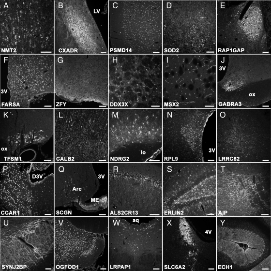
A need exists for mapping the protein profiles in the human brain both during normal and disease conditions. Here we studied 800 antibodies generated toward human proteins as part of a Human Protein Atlas program and investigated their suitability for detailed analysis of various levels of a rat brain using immuno-based methods. In this way, the parallel, rather limited analysis of the human brain, restricted to four brain areas (cerebellum, cerebral cortex, hippocampus, and lateral subventricular zone), could be extended in the rat model to 25 selected areas of the brain. Approximately 100 antibodies (12%) revealed a distinct staining pattern and passed validation of specificity using Western blot analysis. These antibodies were applied to coronal sections of the rat brain at 0.7-mm intervals covering the entire brain. We have now produced detailed protein distribution profiles for these antibodies and acquired over 640 images that form the basis of a publicly available portal of an antibody-based Rodent Brain Protein Atlas database (www.proteinatlas.org/rodentbrain). Because of the systematic selection of target genes, the majority of antibodies included in this database are generated against proteins that have not been studied in the brain before. Furthermore optimized tissue processing and colchicine treatment allow a high quality, more extended annotation and detailed analysis of subcellular distributions and protein dynamics.


Figures

Fig. 1.
Fig. 1.
Schematic overview of the 25 selected brain areas. Included are telencephalon (medial septum, lateral septum, horizontal/vertical diagonal band, prefrontal/cingulate/somatosensory/piriform/entorhinal cortex, ventral pallidum, stria terminalis, globus pallidus, caudate putamen, amygdala (basolateral, central, and medial), hippocampus, and dentate gyrus); diencephalon (preoptic area (A), supraoptic nucleus (A), suprachiasmatic nucleus (A), paraventricular nucleus (A and B), arcuate nucleus (B), median eminence (B), and thalamus); mesencephalon (substantia nigra, ventral tegmental area, and raphe nucleus (dorsal and median)); pons (locus caeruleus (C)); and cerebellum.
Fig. 2.
Fig. 2.
Western blot results of all 32 antibodies used in the study. Left column, protein ladder (green); middle column, Western blot result (red); right column, Western blot result (red) and theoretical band of predicted size ±20% or +10 kDa (yellow) based on prediction (Ensembl database). See Table I for protein names.
Fig. 3.
Fig. 3.
Immunofluorescence micrographs showing a spectrum of staining patterns of 25 validated antibodies in 25 selected, different brain areas. Antibody HPA001303 (NMT2) stains the soma of neurons in many regions, including medial septum (A). HPA003342 (CXADR) stains nerve terminals in many areas including lateral septum (B). HPA002114 (PSMD14) visualizes a nuclear localization mainly in widespread neurons, including cingulate cortex (C). HPA001814 (SOD2) reveals a dotted staining of cell soma in many brain areas, including ventral pallidum (D). HPA001922 (RAP1GAP) stains many neuron subpopulations, labeling soma, dendrites, and axons in many brain areas, including stria terminalis (E). HPA001911 (FARSA) stains many neurons in many regions, including the preoptic area, with immunoreactivity being limited to the cell soma (F). HPA000723 (ZFY) reveals a dotted staining pattern in many brain areas, including the horizontal and vertical diagonal band nuclei, and is strongest around blood vessels (G). HPA001648 (DDX3X) stains many neuronal cell bodies (possibly also some non-neuronal cells) in the brain, including globus pallidus (H). HPA005652 (MSX2) stains many blood vessels in the rat CNS, including in caudate putamen (I). HPA000839 (GABRA3) stains nerve terminals in many areas of the brain including the suprachiasmatic nucleus (J). HPA000337 (TFSM1) stains neurons in the paraventricular and supraoptic nucleus after colchicine treatment (K). HPA007306 (CALB2) stains subsets of neurons and their dendrites and axons throughout the brain, including somatosensory cortex (L). HPA002896 (NDRG2) gives an astrocyte-like staining pattern throughout the brain, including piriform cortex (M). HPA003372 (RPL9) stains many neuronal and non-neuronal cells throughout the brain, including ependymal cells along the third ventricle wall and the paraventricular nucleus (N). HPA000781 (LRRC62) stains axons in various brain regions, including the amygdaloid complex (O). HPA007856 (CCAR1) stains nuclei of neurons and non-neuronal cells throughout the CNS, including the thalamus (P). HPA006641 (SCGN) stains neurons in a few brain areas, including the arcuate nucleus, also axons and/or non-neuronal cells in the median eminence and other brain regions (Q). HPA003229 (ALSCR13) reveals a dendritic and/or nerve terminal staining pattern in many brain areas, including the hippocampus (CA1) with variable intensity (R). HPA002025 (ERLIN2) gives a dotted staining and shows immunoreactivity both in neurons and non-neuronal cells in many brain areas, including dentate gyrus (S). HPA004063 (AIP) stains neurons and their axons and nerve terminals in various areas, including the ventral tegmental area (T). HPA000866 (SYNJ2BP) stains many neurons and their nerve terminals throughout the rat CNS; in the area containing the substantia nigra cell bodies and proximal dendrites are stained (U). HPA003215 (OGFOD1) shows a widespread nuclear staining, including in neurons in the entorhinal cortex (V). HPA008001 (LRPAP1) stains subsets of neurons and dendrites in various brain areas, including the dorsal raphe nucleus (W). HPA004057 (SLC6A2) strongly stains noradrenaline neurons in the locus caeruleus and the axons of these cells throughout the CNS (X). HPA005835 (ECH1) labels non-neuronal cells throughout the rat CNS including Bergmann radial glia in the cerebellum. For gene names and information see Table I. 3V, third ventricle; aq, aqueduct; Arc, arcuate nucleus; D3V, dorsal third ventricle; lo, lateral olfactory tract; LV, lateral ventricle; ME, median eminence; ox, optic chiasm; 4V, fourth ventricle. Scale bars, 100 μm.
Fig. 4.
Fig. 4.
Distribution of immunoreactivity beyond the 25 selected regions. Antisera producing strong staining included HPA005551 (CFH) showing dendrite/radial glia-like structures in cortical areas and hippocampal formation but also glia cells in the subfornical organ (SFO) (A). HPA000612 (APOOL) labels sparsely distributed fibers throughout the rat CNS; after colchicine treatment neurons can be visualized in the dorsal endopiriform nucleus and posterior hypothalamus (B). HPA002317 (BIRC3) labels axons and nerve terminals in many brain areas but also strongly labeled the subventricular zone (SVZ) of the caudate nucleus (C). HPA001032 (PDCD4) strongly labels cells at the ventral border of the brain and around the recess of third ventricle (D). 3V, third ventricle; cc, corpus callosum; CPu, caudate putamen; LV, lateral ventricle; mt, mammillothalamic tract. Scale bars, 100 |gmm. See Table I for protein names.
Fig. 5.
Fig. 5.
Double labeling experiments confirm labeling of cell types known to express target proteins. HPA005753 is raised against a mitotic kinase (PBK) known to be expressed in neuronal progenitors. Double labeling with the early neuronal marker DCx and the nuclear progenitor marker Ki67 reveals limited co-expression of PBK with DCx (A) but a strong coexistence of PBK with Ki67, indicating the expression of this protein in proliferating cells (B). PBK is localized in the cytoplasmic domain of progenitor cells (B, arrowheads). A three-dimensional reconstruction of a progenitor cell in the subventricular zone (C) reveals the distribution of PBK in the cytosol, including filopodia/lamellipodia (note the exclusion of PBK from the nucleus). HPA007306 was raised against calretinin (CALB2), a well known marker for subsets of cortical interneurons. When applied to sections of a GAD67-GFP transgenic mouse, this antibody labels a subset of GABAergic interneurons (E, arrows) in the somatosensory cortex (D and E). cc, corpus callosum; LV, lateral ventricle. Scale bars are: A and D, 50 μm; B, 25 μm; C, 10 μm; E, 25 μm.
Fig. 6.
Fig. 6.
HPA007959 (C6orf64), raised against a protein of unknown function located on chromosome 6, labeled neurons in the paraventricular (A and B) and supraoptic (C and D) nuclei and their projections. Co-labeling with vasopressin (AVP) (A and C) and oxytocin (OT) (B and D) reveals immunoreactivity exclusively in oxytocin-producing neurons. Scale bars in A–D, 100 μm. ox, optic chiasm; 3V, third ventricle.
Fig. 7.
Fig. 7.
Colchicine treatment prevents centrifugal transport, allowing analysis of protein dynamics and identification of cells producing these proteins. Antibody HPA003229 (ALS2CR13) reveals a synaptic staining in layer Ib of the piriform cortex (A). In brain tissue treated with colchicine ALS2CR13 is located in proximal dendrites (B). Antibody HPA001830 (SNAP25) strongly stains synapses in the somatosensory cortex (C). When applied to brain sections of colchicine-treated rats, a subset of neurons in cortex shows immunoreactivity (D). This could indicate postsynaptic localization of ALS2CR13 and presynaptic localization of SNAP25. I–III denote the different layers of the piriform cortex; lo, lateral olfactory tract. Scale bars, 100 μm.

Similar articles

Cited by

References

    1. Lein E. S., Hawrylycz M. J., Ao N., Ayres M., Bensinger A., Bernard A., Boe A. F., Boguski M. S., Brockway K. S., Byrnes E. J., Chen L., Chen L., Chen T. M., Chin M. C., Chong J., Crook B. E., Czaplinska A., Dang C. N., Datta S., Dee N. R., Desaki A. L., Desta T., Diep E., Dolbeare T. A., Donelan M. J., Dong H. W., Dougherty J. G., Duncan B. J., Ebbert A. J., Eichele G., Estin L. K., Faber C., Facer B. A., Fields R., Fischer S. R., Fliss T. P., Frensley C., Gates S. N., Glattfelder K. J., Halverson K. R., Hart M. R., Hohmann J. G., Howell M. P., Jeung D. P., Johnson R. A., Karr P. T., Kawal R., Kidney J. M., Knapik R. H., Kuan C. L., Lake J. H., Laramee A. R., Larsen K. D., Lau C., Lemon T. A., Liang A. J., Liu Y., Luong L. T., Michaels J., Morgan J. J., Morgan R. J., Mortrud M. T., Mosqueda N. F., Ng L. L., Ng R., Orta G. J., Overly C. C., Pak T. H., Parry S. E., Pathak S. D., Pearson O. C., Puchalski R. B., Riley Z. L., Rockett H. R., Rowland S. A., Royall J. J., Ruiz M. J., Sarno N. R., Schaffnit K., Shapovalova N. V., Sivisay T., Slaughterbeck C. R., Smith S. C., Smith K. A., Smith B. I., Sodt A. J., Stewart N. N., Stumpf K. R., Sunkin S. M., Sutram M., Tam A., Teemer C. D., Thaller C., Thompson C. L., Varnam L. R., Visel A., Whitlock R. M., Wohnoutka P. E., Wolkey C. K., Wong V. Y., Wood M., Yaylaoglu M. B., Young R. C., Youngstrom B. L., Yuan X. F., Zhang B., Zwingman T. A., Jones A. R. ( 2007) Genome-wide atlas of gene expression in the adult mouse brain. Nature 445, 168– 176 - PubMed
    1. Sandberg R., Yasuda R., Pankratz D. G., Carter T. A., Del Rio J. A., Wodicka L., Mayford M., Lockhart D. J., Barlow C. ( 2000) Regional and strain-specific gene expression mapping in the adult mouse brain. Proc. Natl. Acad. Sci. U.S.A. 97, 11038– 11043 - PMC - PubMed
    1. Liao B. Y., Zhang J. ( 2006) Evolutionary conservation of expression profiles between human and mouse orthologous genes. Mol. Biol. Evol. 23, 530– 540 - PubMed
    1. Premack D. ( 2007) Human and animal cognition: continuity and discontinuity. Proc. Natl. Acad. Sci. U.S.A. 104, 13861– 13867 - PMC - PubMed
    1. Gibbs R. A., Weinstock G. M., Metzker M. L., Muzny D. M., Sodergren E. J., Scherer S., Scott G., Steffen D., Worley K. C., Burch P. E., Okwuonu G., Hines S., Lewis L., DeRamo C., Delgado O., Dugan-Rocha S., Miner G., Morgan M., Hawes A., Gill R., Celera Holt R. A., Adams M. D., Amanatides P. G., Baden-Tillson H., Barnstead M., Chin S., Evans C. A., Ferriera S., Fosler C., Glodek A., Gu Z., Jennings D., Kraft C. L., Nguyen T., Pfannkoch C. M., Sitter C., Sutton G. G., Venter J. C., Woodage T., Smith D., Lee H. M., Gustafson E., Cahill P., Kana A., Doucette-Stamm L., Weinstock K., Fechtel K., Weiss R. B., Dunn D. M., Green E. D., Blakesley R. W., Bouffard G. G., De, Jong P. J., Osoegawa K., Zhu B., Marra M., Schein J., Bosdet I., Fjell C., Jones S., Krzywinski M., Mathewson C., Siddiqui A., Wye N., McPherson J., Zhao S., Fraser C. M., Shetty J., Shatsman S., Geer K., Chen Y., Abramzon S., Nierman W. C., Havlak P. H., Chen R., Durbin K. J., Egan A., Ren Y., Song X. Z., Li B., Liu Y., Qin X., Cawley S., Worley K. C., Cooney A. J., D'Souza L. M., Martin K., Wu J. Q., Gonzalez-Garay M. L., Jackson A. R., Kalafus K. J., McLeod M. P., Milosavljevic A., Virk D., Volkov A., Wheeler D. A., Zhang Z., Bailey J. A., Eichler E. E., Tuzun E., Birney E., Mongin E., Ureta-Vidal A., Woodwark C., Zdobnov E., Bork P., Suyama M., Torrents D., Alexandersson M., Trask B. J., Young J. M., Huang H., Wang H., Xing H., Daniels S., Gietzen D., Schmidt J., Stevens K., Vitt U., Wingrove J., Camara F., Mar Albà M., Abril J. F., Guigo R., Smit A., Dubchak I., Rubin E. M., Couronne O., Poliakov A., Hübner N., Ganten D., Goesele C., Hummel O., Kreitler T., Lee Y. A., Monti J., Schulz H., Zimdahl H., Himmelbauer H., Lehrach H., Jacob H. J., Bromberg S., Gullings-Handley J., Jensen-Seaman M. I., Kwitek A. E., Lazar J., Pasko D., Tonellato P. J., Twigger S., Ponting C. P., Duarte J. M., Rice S., Goodstadt L., Beatson S. A., Emes R. D., Winter E. E., Webber C., Brandt P., Nyakatura G., Adetobi M., Chiaromonte F., Elnitski L., Eswara P., Hardison R. C., Hou M., Kolbe D., Makova K., Miller W., Nekrutenko A., Riemer C., Schwartz S., Taylor J., Yang S., Zhang Y., Lindpaintner K., Andrews T. D., Caccamo M., Clamp M., Clarke L., Curwen V., Durbin R., Eyras E., Searle S. M., Cooper G. M., Batzoglou S., Brudno M., Sidow A., Stone E. A., Venter J. C., Payseur B. A., Bourque G., López-Otín C., Puente X. S., Chakrabarti K., Chatterji S., Dewey C., Pachter L., Bray N., Yap V. B., Caspi A., Tesler G., Pevzner P. A., Haussler D., Roskin K. M., Baertsch R., Clawson H., Furey T. S., Hinrichs A. S., Karolchik D., Kent W. J., Rosenbloom K. R., Trumbower H., Weirauch M., Cooper D. N., Stenson P. D., Ma B., Brent M., Arumugam M., Shteynberg D., Copley R. R., Taylor M. S., Riethman H., Mudunuri U., Peterson J., Guyer M., Felsenfeld A., Old S., Mockrin S., Collins F. ( 2004) Genome sequence of the Brown Norway rat yields insights into mammalian evolution. Nature 428, 493– 521 - PubMed

Publication types

LinkOut - more resources