. 2009 May 1;122(Pt 9):1315-21.
doi: 10.1242/jcs.043216. Epub 2009 Apr 7.

Copper transport into the secretory pathway is regulated by oxygen in macrophages

Affiliations

Copper transport into the secretory pathway is regulated by oxygen in macrophages

Carine White et al. J Cell Sci. .

Abstract

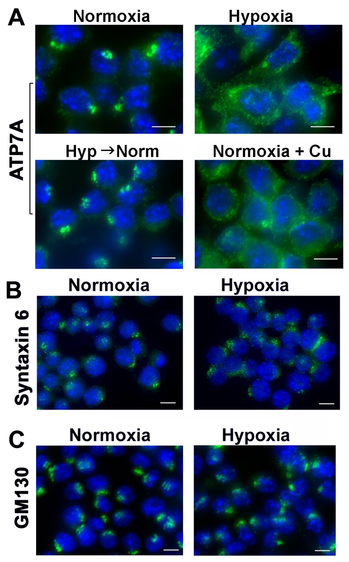
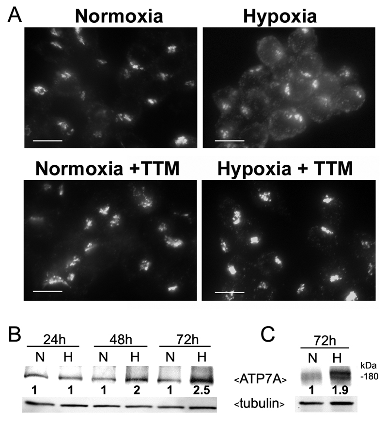
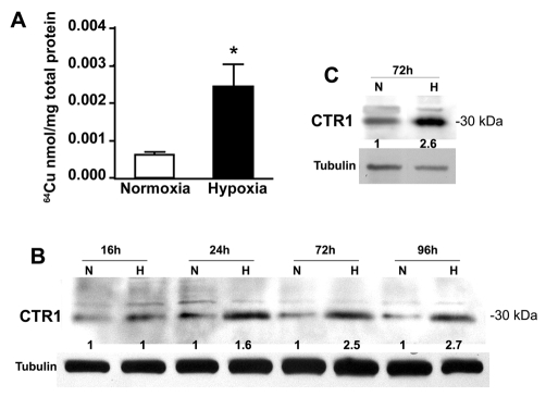
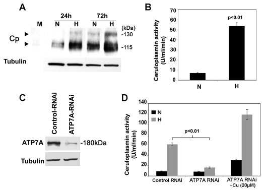
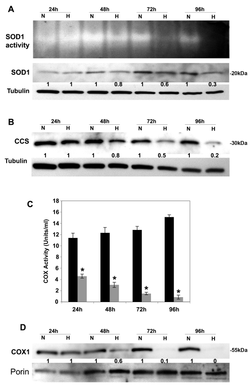
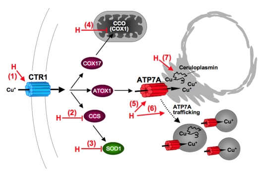
Copper is an essential nutrient for a variety of biochemical processes; however, the redox properties of copper also make it potentially toxic in the free form. Consequently, the uptake and intracellular distribution of this metal is strictly regulated. This raises the issue of whether specific pathophysiological conditions can promote adaptive changes in intracellular copper distribution. In this study, we demonstrate that oxygen limitation promotes a series of striking alterations in copper homeostasis in RAW264.7 macrophage cells. Hypoxia was found to stimulate copper uptake and to increase the expression of the copper importer, CTR1. This resulted in increased copper delivery to the ATP7A copper transporter and copper-dependent trafficking of ATP7A to cytoplasmic vesicles. Significantly, the ATP7A protein was required to deliver copper into the secretory pathway to ceruloplasmin, a secreted copperdependent enzyme, the expression and activity of which were stimulated by hypoxia. However, the activities of the alternative targets of intracellular copper delivery, superoxide dismutase and cytochrome c oxidase, were markedly reduced in response to hypoxia. Collectively, these findings demonstrate that copper delivery into the biosynthetic secretory pathway is regulated by oxygen availability in macrophages by a selective increase in copper transport involving ATP7A.


Figures

Fig. 1.
Fig. 1.
Hypoxia stimulates trafficking of the ATP7A protein. (A) Immunofluorescence analysis of ATP7A protein in RAW264.7 cells grown under normoxic (21% O2) or hypoxic (4% O2) conditions for 96 hours. Cells were fixed, permeabilized and probed with antibodies against for ATP7A and anti-rabbit antibodies conjugated to Alexa Fluor 488 (green). Nuclei were labeled with DAPI (blue). Note the trafficking of ATP7A protein from the perinuclear region in hypoxic cells and copper-treated normoxic cells. ATP7A was rapidly retrieved to the perinuclear compartment upon the transfer of hypoxic cells to normoxic media for 30 minutes (Hyp→Norm). (B,C) Analysis of Golgi marker proteins in hypoxic RAW264.7 cells. Cells were cultured under hypoxic or normoxic conditions as described in A and probed using antibodies against the trans-Golgi network marker protein syntaxin 6 (B), or the Golgi matrix protein, GM130 (C). Nuclei were labeled with DAPI (blue). Scale bars: 7 μm.
Fig. 2.
Fig. 2.
Hypoxia stimulates both copper-dependent trafficking and increased expression of ATP7A. (A) RAW264.7 cells were cultured under hypoxic or normoxic conditions as described in Fig. 1 in the presence or absence of the copper chelator, tetrathiomolybdate (TTM; 5 nM). ATP7A protein was detected using immunofluorescence as described in Fig. 1. Scale bars: 10 μm. (B,C) Immunoblot analysis of ATP7A protein in RAW264.7 cells (B) and primary peritoneal macrophages (C) cultured under normoxic (N; 21% O2) or hypoxic (H; 4% O2) conditions for the indicated times. Tubulin was detected as a loading control. Relative ATP7A band intensities at each time point normalized against tubulin are shown for each normoxic and hypoxic pair.
Fig. 3.
Fig. 3.
ATP7A trafficking in HIF1α-positive tumor-associated macrophages. (A) ATP7A is strongly expressed in tumor-associated macrophages. Human prostate cell PC-3 xenograft tumors from SCID mice were cryosectioned and probed with antibodies against ATP7A (green) or antibodies against the macrophage marker CD68 (red). Nuclei were stained using DAPI (blue). Upper panels show both the tumor mass and the tumor edge, with strong ATP7A expression in CD68-positive macrophages associated with the tumor edge Scale bar: 60 μm. The lower panels are higher magnifications of the tumor periphery and reveal extensive coexpression of ATP7A in CD68-positive macrophages, as indicated by yellow signal in the merged image. Scale bar: 30 μm. The arrow and arrowhead (lower left panel) indicate heterogeneous localization of the ATP7A in macrophages, in either a perinuclear or dispersed distribution. (B) The dispersed distribution of the ATP7A occurs in HIF1α-positive macrophages. Three different PC-3 tumor cryosections were immunostained for ATP7A (green) and HIF1α staining (red). ATP7A was restricted to the perinuclear region of macrophages negative for HIF1α expression (arrows), whereas a dispersed distribution of ATP7A occurred in HIF1α-positive macrophages (arrowheads) (original magnification, ×600). Scale bar: 10 μm.
Fig. 4.
Fig. 4.
Hypoxia stimulates copper uptake and CTR1 expression in RAW264.7 macrophages. (A) Copper uptake activity. RAW264.7 cells were pre-exposed to normoxia (21% O2) or hypoxia (4% O2) for 72 hours and 64Cu uptake was measured over 5 minutes. Values were normalized against total protein concentrations (mean + s.d.; n=3; *P<0.05). (B,C) The effect of hypoxia on CTR1 protein levels in RAW264.7 cells (B) and primary peritoneal macrophages (C) cultured under normoxia (N; 21% O2) or hypoxia (H; 4% O2) for the indicated times. Immunoblot analysis was used to detect CTR1 protein in lysates using anti-CTR1 antibodies. Tubulin was detected as a loading control. Relative CTR1 band intensities at each time point, normalized against tubulin, are shown for each normoxic and hypoxic pair.
Fig. 5.
Fig. 5.
ATP7A-dependent copper transport is required for hypoxia-stimulated ceruloplasmin activity. (A) Immunoblot analysis of ceruloplasmin (Cp) secreted from RAW264.7 cells grown under normoxic (N; 21% O2) or hypoxic (H; 4% O2) conditions for the indicated times. Conditioned medium was concentrated and subjected to non-denaturing SDS-PAGE and immunoblot analysis with anti-Cp antibodies. Tubulin levels from corresponding cell lysates are also shown. Lane 1 is a negative control of concentrated growth medium alone (M). (B) Hypoxia stimulates ceruloplasmin activity. Ceruloplasmin activity (p-phenylenediamine oxidase activity) was measured in the concentrated conditioned medium from RAW264.7 cells following exposure to normoxia (N; 21% O2) or hypoxia (H; 4% O2) for 72 hours. Activity was normalized against total protein content of the corresponding cell lysates (mean + s.d.; n=3). (C) RNAi-mediated silencing of the ATP7A protein. Immunoblot analysis of ATP7A protein levels in RAW264.7 cells stably transfected with either ATP7A-RNAi or control-RNAi. (D) Ceruloplasmin activity was measured in conditioned medium from the ATP7A-RNAi or control-RNAi cells exposed to hypoxia (N; 21% O2) or hypoxia (H; 4% O2) for 72 hours (mean + s.d.; n=3). Note the failure to activate ceruloplasmin in ATP7A-depleted cells, and the restoration by addition of copper to the growth medium (+Cu).
Fig. 6.
Fig. 6.
Hypoxia downregulates alternative copper pathways in RAW264.7 macrophages. (A) Effect of hypoxia on SOD1 protein and activity. RAW264.7 cells were grown under normoxic (N; 21% O2) or hypoxic (H; 4% O2) conditions for the indicated times. Cell lysates were subjected to non-denaturing SDS-PAGE for the in-gel SOD1 activity assay (top panel). Immunoblots from the same lysates were probed with anti-SOD1 antibodies to detect SOD1 protein. Tubulin was detected as a loading control. Relative SOD1 band intensities at each time point, normalized against tubulin, are shown for each normoxic and hypoxic pair. (B) Effect of hypoxia on the abundance of CCS, the copper chaperone for SOD1. The same lysates as in A were subjected to SDS-PAGE and immunoblot analysis with anti-CCS antibodies and relative band intensities at each time point normalized against tubulin are shown for each normoxic and hypoxic pair. (C) Analysis of CCO activity in hypoxic (shaded bars) and normoxic (solid bars) conditions. Activity was measured in mitochondrial preparations isolated from RAW264.7 cells cultured as described in A. Values were normalized against total mitochondrial protein (mean + s.d.; n=3; *P<0.05). (D) Analysis of COX1 protein levels in RAW264.7 cells cultured under normoxic (N; 21% O2) or hypoxic (H; 4% O2) conditions for the indicated times. Mitochondrial preparations from B were subjected to SDS-PAGE and probed with antibodies against the copper-binding subunit COX1 of the CCO complex. Immunoblots were probed with an antibody against porin to indicate protein loading, and relative COX1 band intensities at each time point, normalized against porin, are shown for each normoxic and hypoxic pair.
Fig. 7.
Fig. 7.
Schematic model of hypoxia-induced changes in copper homeostasis resulting in ATP7A-mediated copper transport to the secretory pathway. Effects of hypoxia (H) include: (1) increased expression of the CTR1 copper importer and increased copper uptake; (2) decreased CCS expression; (3) reduced activity of SOD1; (4) reduced CCO activity and reduced expression of COX1; (5) increased expression of ATP7A; (6) copper-dependent trafficking of ATP7A; and (7) ATP7A-dependent copper transport to the secretory pathway of ceruloplasmin.

References

    1. Alessandri, G., Raju, K. and Gullino, P. M. (1984). Angiogenesis in vivo and selective mobilization of capillary endothelium in vitro by heparin-copper complex. Microcirc. Endothelium Lymphatics 1, 329-346. - PubMed
    1. Amaravadi, R., Glerum, D. M. and Tzagoloff, A. (1997). Isolation of a cDNA encoding the human homolog of COX17, a yeast gene essential for mitochondrial copper recruitment. Hum. Genet. 99, 329-333. - PubMed
    1. Arnold, F., West, D. and Kumar, S. (1987). Wound healing: the effect of macrophage and tumour derived angiogenesis factors on skin graft vascularization. B. J. Exp. Pathol. 68, 569-574. - PMC - PubMed
    1. Barnes, N., Tsivkovskii, R., Tsivkovskaia, N. and Lutsenko, S. (2005). The copper-transporting ATPases, menkes and wilson disease proteins, have distinct roles in adult and developing cerebellum. J. Biol. Chem. 280, 9640-9645. - PubMed
    1. Biswas, S. K., Gangi, L., Paul, S., Schioppa, T., Saccani, A., Sironi, M., Bottazzi, B., Doni, A., Vincenzo, B., Pasqualini, F. et al. (2006). A distinct and unique transcriptional program expressed by tumor-associated macrophages (defective NF-kappaB and enhanced IRF-3/STAT1 activation). Blood 107, 2112-2122. - PubMed

Publication types