. 2009 Apr 15;69(8):3256-61.
doi: 10.1158/0008-5472.CAN-08-4055. Epub 2009 Apr 7.

PTEN loss contributes to erlotinib resistance in EGFR-mutant lung cancer by activation of Akt and EGFR

Affiliations

PTEN loss contributes to erlotinib resistance in EGFR-mutant lung cancer by activation of Akt and EGFR

Martin L Sos et al. Cancer Res. .

Erratum in

Abstract

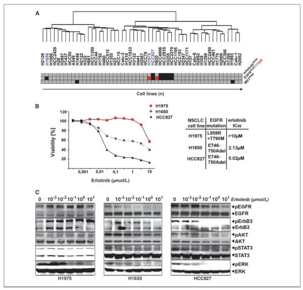
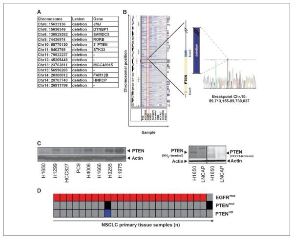
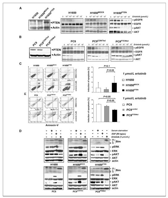
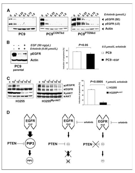
Clinical resistance to epidermal growth factor receptor (EGFR) inhibition in lung cancer has been linked to the emergence of the EGFR T790M resistance mutation or amplification of MET. Additional mechanisms contributing to EGFR inhibitor resistance remain elusive. By applying combined analyses of gene expression, copy number, and biochemical analyses of EGFR inhibitor responsiveness, we identified homozygous loss of PTEN to segregate EGFR-dependent and EGFR-independent cells. We show that in EGFR-dependent cells, PTEN loss partially uncouples mutant EGFR from downstream signaling and activates EGFR, thereby contributing to erlotinib resistance. The clinical relevance of our findings is supported by the observation of PTEN loss in 1 out of 24 primary EGFR-mutant non-small cell lung cancer (NSCLC) tumors. These results suggest a novel resistance mechanism in EGFR-mutant NSCLC involving PTEN loss.


Figures

Figure 1
Figure 1
An EGFR independence signature in H1650 cells. A, hierarchical clustering of 53 NSCLC cells according to gene expression. Erlotinib sensitivity (IC50 < 1 μmol/L, red; IC50 > 1 μmol/L, gray) and EGFR mutations (EGFR-mutant, black; T790M, red; EGFR wild-type, gray) as well as MET amplification (black). B, left, cellular viability as a function of erlotinib dose for all three cell lines studied. Right, mutation status and IC50 values. C, cells were treated with different doses of erlotinib. Activation of EGFR and downstream signaling pathways was determined by analyzing the amount of phosphorylated versions of the respective proteins in comparison with their total levels using phosphorylation-specific antibodies.
Figure 2
Figure 2
Genomic characterization of PTEN loss in H1650 cells. A, list of genes affected by differential lesions between H1650 cells and EGFR-mutant and erlotinib-sensitive cell lines. B, left, screenshot showing chromosomal aberrations at chromosome 10 (Integrative Genomics Viewer; http://www.broad.mit.edu/igv/) of all EGFR-mutant cells. Middle, 3′-region mapping of PTEN using quantitative PCR reveals a homozygous deletion deleting parts of exon 8 and the entire exon 9. Right, the sequence bridging the breakpoint. C, left, PTEN protein status determined using immunoblotting in different NSCLC cell lines. Right, NH2-terminal and COOH-terminal PTEN detection by immunoblotting. LNCAP cells, known to express a truncated version of PTEN, served as controls. D, analysis of EGFR mutations (red) and homozygous deletions of PTEN (black) and PTEN mutations (blue) in 140 lung cancer biopsy specimens.
Figure 3
Figure 3
Erlotinib resistance in EGFR-mutated NSCLC with PTEN loss. A, left, in H1650PTEN cells, PTEN levels were determined by immunoblotting. Right, levels of phospho-EGFR and phospho-AKT were assessed by immunoblotting in H1650, H1650MOCK, and H1650PTEN cells treated with erlotinib. B, left, in PC9PTENkd cells, PTEN levels were determined by immunoblotting. Right, levels of phospho-EGFR and phospho-AKT were assessed in PC9, PC9CONTkd, and PC9PTENkd cells treated with erlotinib. C, left, percentage of apoptotic cells (in %, analyzed by measuring the fraction of cells positive for Annexin V and/or propidium iodide by flow cytometry) after treatment with either erlotinib (1 μmol/L) or control. Right, cumulative histograms of apoptosis induction. D, levels of Bim (EL, extra long; L, long; S, short), phospho-ERK, phospho-pAKT, and actin were measured after serum starvation (serum starvation “+”), EGF stimulation (EGF “+”), or treatment with erlotinib (1 μmol/L erlotinib “+”) for 24 h.
Figure 4
Figure 4
PTEN loss activates EGFR. A, phospho-EGFR was detected by immunoblotting after short exposure (SE) and long exposure (LE) in PC9, PC9CONTkd, and PC9PTENkd cells. Actin levels served as a loading control. B, left, levels of phospho-EGFR of PC9PTENkd and PC9 cells treated with erlotinib were determined (+/− EGF) under serum starvation. Right, apoptosis (%) after erlotinib treatment (0.5 μmol/L) in the given cells. C, left, phospho-EGFR and phospho-AKT in H3255 and H3255MyrAKT cells were assessed by immunoblotting. Right, the fraction of apoptotic cells (in %) in the given cells. D, a simplified model explaining our observations: in EGFR-mutant cells, EGFR is the sole input for production of PIP3. Inhibiting EGFR dramatically reduces the input into PIP3 production. Therefore, the lack of negative regulation of PIP3 production by loss of PTEN is limited.

References

    1. Sharma SV, Fischbach MA, Haber DA, Settleman J. “Oncogenic shock”: explaining oncogene addiction through differential signal attenuation. Clin Cancer Res. 2006;12:4392–5s. - PubMed
    1. Thomas RK, Greulich H, Yuza Y, et al. Detection of oncogenic mutations in the EGFR gene in lung adenocarcinoma with differential sensitivity to EGFR tyrosine kinase inhibitors. Cold Spring Harb Symp Quant Biol. 2005;70:73–81. - PubMed
    1. Engelman JA, Janne PA. Mechanisms of acquired resistance to epidermal growth factor receptor tyrosine kinase inhibitors in non-small cell lung cancer. Clin Cancer Res. 2008;14:2895–9. - PubMed
    1. Bean J, Brennan C, Shih JY, et al. MET amplification occurs with or without T790M mutations in EGFR mutant lung tumors with acquired resistance to gefitinib or erlotinib. Proc Natl Acad Sci U S A. 2007;104:20932–7. - PMC - PubMed
    1. Guo A, Villen J, Kornhauser J, et al. Signaling networks assembled by oncogenic EGFR and c-Met. Proc Natl Acad Sci U S A. 2008;105:692–7. - PMC - PubMed

Publication types

MeSH terms