. 2009 Apr 15;69(8):3472-81.
doi: 10.1158/0008-5472.CAN-08-3886. Epub 2009 Apr 7.

Human glioblastoma-derived cancer stem cells: establishment of invasive glioma models and treatment with oncolytic herpes simplex virus vectors

Affiliations

Human glioblastoma-derived cancer stem cells: establishment of invasive glioma models and treatment with oncolytic herpes simplex virus vectors

Hiroaki Wakimoto et al. Cancer Res. .

Abstract

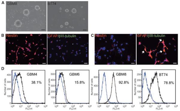
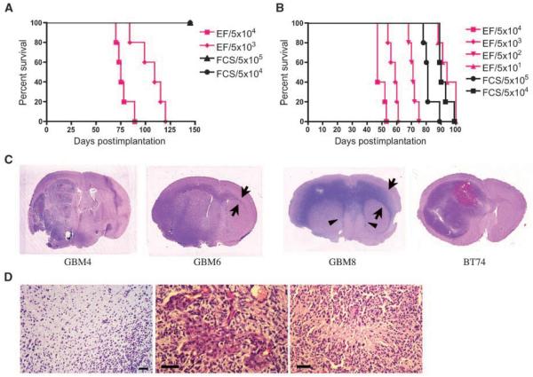
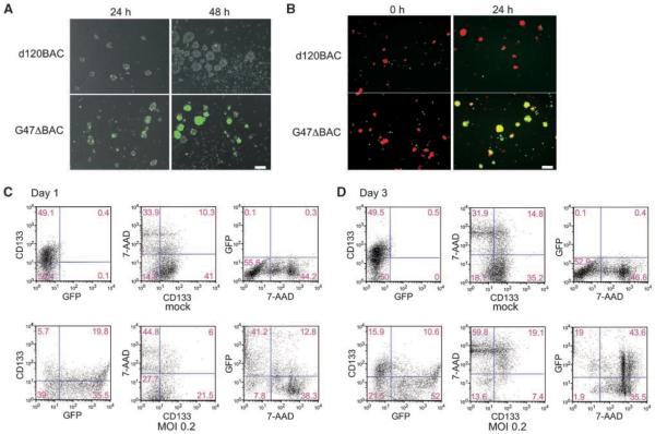
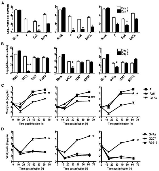
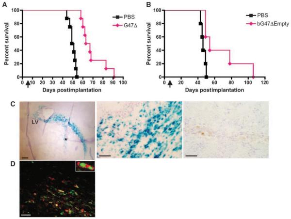
Glioblastoma, the most malignant type of primary brain tumor, is one of the solid cancers where cancer stem cells have been isolated, and studies have suggested resistance of those cells to chemotherapy and radiotherapy. Here, we report the establishment of CSC-enriched cultures derived from human glioblastoma specimens. They grew as neurospheres in serum-free medium with epidermal growth factor and fibroblast growth factor 2, varied in the level of CD133 expression and very efficiently formed highly invasive and/or vascular tumors upon intracerebral implantation into immunodeficient mice. As a novel therapeutic strategy for glioblastoma-derived cancer stem-like cells (GBM-SC), we have tested oncolytic herpes simplex virus (oHSV) vectors. We show that although ICP6 (UL39)-deleted mutants kill GBM-SCs as efficiently as wild-type HSV, the deletion of gamma34.5 significantly attenuated the vectors due to poor replication. However, this was significantly reversed by the additional deletion of alpha47. Infection with oHSV G47Delta (ICP6(-), gamma34.5(-), alpha47(-)) not only killed GBM-SCs but also inhibited their self-renewal as evidenced by the inability of viable cells to form secondary tumor spheres. Importantly, despite the highly invasive nature of the intracerebral tumors generated by GBM-SCs, intratumoral injection of G47Delta significantly prolonged survival. These results for the first time show the efficacy of oHSV against human GBM-SCs, and correlate this cytotoxic property with specific oHSV mutations. This is important for designing new oHSV vectors and clinical trials. Moreover, the new glioma models described in this study provide powerful tools for testing experimental therapeutics and studying invasion and angiogenesis.


Figures

Figure 1
Figure 1
In vitro characterization of human GBM-derived cancer stem cells. A, typical appearance of “neurosphere” structures grown in the serum-free specific culture conditions (GBM8, left; BT74, right). Scale bars, 100 μm. B and C, immunocytochemical analysis of human GBM-SCs and their differentiated progeny. B, undifferentiated BT74 cells; C, differentiated BT74 cells. Left, staining for neural stem/progenitor marker nestin (Cy3, red); right, merged images of staining for astrocytic marker GFAP (Cy3, red) and neuronal marker βIII tubulin (FITC, green). A significant reduction in nestin expression and concomitant up-regulation of GFAP expression were observed after induction of differentiation. There are cells double positive for GFAP and βIII tubulin in the differentiated population (yellow, white arrows). Nuclei were visualized by staining with 4′,6-diamidino-2-phenylindole. Scale bars, 20 μm. D, flow cytometric analysis for neuronal stem cell marker CD133 on GBM-SC cultures. Dissociated neurospheres were stained either with isotype control (blue line) or anti-CD133/2 antibody (black line) conjugated with phycoerythrin before the analysis. The percentage of CD133-positive cells is indicated.
Figure 2
Figure 2
In vivo tumorigenicity of GBM-SCs and histopathology of the xenografts. A and B, Kaplan-Meier survival curves of mice implanted with GBM4 cells (A) or GBM8 cells (B). Immunodeficient mice were orthotopically implanted with cells grown in EF medium (pink lines), or in 10% FCS medium (FCS, black lines). Numbers, cell number implanted. Mice were followed for survival. n = 5 mice per group. Median survival times were statistically different between groups that received 5 × 10⁴ EF cells and FCS cells (in both GBM4 and GBM8 cells; logrank test, P < 0.0001). C, photographs of magnified coronal sections of xenografts stained with H&E. GBM4 tumor was characterized by a massive, multilobulated, and hypervascular mass with intratumoral bleeding. GBM6 tumor and GBM8 tumor extended through the corpus callosum to the contralateral brain (arrows). GBM8 tumor mimicked the butterfly-like growth pattern characteristic of human GBM, and tended to grow along subventricular areas, compressing the lateral ventricles (arrowheads). BT74 cells formed a massive tumor with intermediate invasiveness and hemorrhage resulting in an enlarged hemisphere. D, GBM-SC-derived intracerebral xenografts recapitulate GBM histopathology. Left, showing tumor cell infiltration into surrounding brain without distinctive border (GBM8). Endovascular proliferation (middle) and pseudopalisading necrosis (right), hallmarks of the GBM histopathology, were observed (BT74). Scale bars, 50 μm.
Figure 3
Figure 3
oHSV is able to infect, replicate, and spread in GBM-SC cultures, killing both CD133-positive and -negative cells. A, growing neurospheres (GBM8) were dissociated to single-cell suspension to ensure interaction of virus with individual cells. Cells were infected with either replication-defective vector d120BAC (top) or replication-competent vector G47ΔBAC (bottom) at MOI of 0.2, and the expression of EGFP (green) was observed serially. At 24 h postinfection, infection with both viral vectors resulted in comparable degrees of EGFP+ cells, whereas at 48 h postinfection, G47ΔBAC, but not d120BAC, showed a large increase in the proportion of EGFP-expressing cells, indicating viral replication and spread. Overlaid images of fluorescence and phase contrast are shown. Scale bar, 100 μm. B, oncolytic viral infection spreads cell-to-cell among GBM-SCs. GBM8 cells were infected with either d120BAC (top) or G47ΔBAC (bottom) at MOI of 1, washed, and plated with uninfected GBM-SCs labeled with orange dye (0 h). The merged images of green and orange fluorescence were recorded 24 h later. G47ΔBAC generated cells double-positive for EGFP and orange fluorescence (yellow) indicative of cell-to-cell infectious spread, whereas d120BAC did not. Scale bar, 100 μm. C and D, flow cytometric analysis of oncolytic viral infection, CD133 status, and cell death. GBM8 cells were either mock-infected (top) or infected with EGFP-expressing G47ΔBAC at MOI of 0.2 (bottom). On days 1 (C) and 3 (D) postinfection, the cells were stained with phycoerythrin-conjugated anti-CD133 antibody, and nonviable cells labeled with 7-ADD, before being subjected to 3-color fluorescence-activated cell sorting analysis. Numbers, percent of cells in each quadrant.
Figure 4
Figure 4
GBM-SCs are susceptible to killing and replication by oHSVs. A and B, oHSVs mediate killing of GBM-SCs, GBM4 (left), GBM8 (middle), and BT74 (right). A, dissociated GBM-SCs were infected with mock, G47Δ, FΔ6, or strain F at MOI of 0.2, and 2 × 10⁴ cells were plated in EF medium in 24-well plates. After 3 and 7 d in culture, trypan blue-excluding viable cells were counted for each well. All the viruses exerted significant cell killing activity against the three tested GBM-SCs, albeit with different potency. FΔ6 kills cells as efficiently as wild-type strain F, whereas G47Δ is less potent than FΔ6 in 2 of 3 GBM-SCs (*, P < 0.05; FΔ6 versus G47Δ). B, comparison of cytotoxicity among three different γ34.5 mutants. G47Δ displays greater cell killing at day 7 compared with G207 and R3616. *, P < 0.05; **, P < 0.01 compared with the values of G207. C and D, replication of oHSVs in GBM-SCs in vitro. GBM4 (left), GBM8 (middle), and BT74 (right) cells were infected as above, and harvested with medium at the indicated time points for virus yield to be determined. C, strain F, FΔ6, and G47Δ all displayed significant viral replication in the tested GBM-SCs, whereas the magnitude of FΔ6 replication was greater than that of G47Δ in all 3 cells (*, P < 0.0001; **, P < 0.005). D, G47Δ displays reasonable viral replication, whereas G207 and R3616 do not, with virus yields much less than input (4,000 pfu per well) over the 62-h time course. *, P < 0.0001, G47Δ compared with G207 or R3616. All experiments were performed in triplicate and data are presented as mean with SD.
Figure 5
Figure 5
Intratumoral injection of G47Δ prolongs survival of mice bearing GBM-SC xenografts. A and B, Kaplan-Meier survival curves of the mice bearing GBM-SCs xenografts treated with oHSV vectors. Forty thousand GBM8EF cells (A) or 2 × 10⁴ BT74 cells (B) were implanted into the brains of athymic (A) or severe combined immunodeficient (B) mice. Six (A) or 7 (B) d later, G47Δ (A) or bG47Δ-Empty (B; 2 × 10⁶ pfu, pink diamond) or PBS (black square) was stereotactically injected at the same coordinates as the tumor cells. Treatment with G47Δ resulted in significantly prolonged survival compared with mock treatment (median survival time, 62 versus 50 d, P < 0.0001 for A; median survival time 54 versus 46 d, P < 0.03 for B). n = 8 (A) or 5 (B) per group. Arrows, time of virus injection. C and D, G47Δ infects GBM-SC tumors in vivo. Twenty-four hours after injection of G47Δ (left and middle in C and D) or PBS (C, right) into GBM8 xenografts, the brains were collected. X-gal staining of the sections (C) revealed an extensive infection of tumor tissue that displays a progression along white matter tracts (left). LV, lateral ventricle. *, injection track. Scale bar, 200 μm. Efficient in vivo infection by G47Δ is shown at higher power magnification (middle), whereas no lacZ positivity is seen in a PBS-treated section (right). Scale bars, 50 μm. D, immunofluorescent staining showing colocalized detection of β-galactosidase (Cy3, red) and human nuclei (FITC, green) on a G47Δ-infected brain section. Scale bar, 50 μm.

References

    1. Wen PY, Kesari S. Malignant gliomas in adults. N Engl J Med. 2008;359:492–507. - PubMed
    1. Dalerba P, Cho RW, Clarke MF. Cancer stem cells: models and concepts. Annu Rev Med. 2007;58:267–84. - PubMed
    1. Galli R, Binda E, Orfanelli U, et al. Isolation and characterization of tumorigenic, stem-like neural precursors from human glioblastoma. Cancer Res. 2004;64:7011–21. - PubMed
    1. Singh SK, Hawkins C, Clarke ID, et al. Identification of human brain tumour initiating cells. Nature. 2004;432:396–401. - PubMed
    1. Yuan X, Curtin J, Xiong Y, et al. Isolation of cancer stem cells from adult glioblastoma multiforme. Oncogene. 2004;23:9392–400. - PubMed

Publication types

MeSH terms