Comparative Study
. 2009 Jul;330(1):283-93.
doi: 10.1124/jpet.109.150672. Epub 2009 Apr 7.

The intracellular II-III loops of Cav1.2 and Cav1.3 uncouple L-type voltage-gated Ca2+ channels from glucagon-like peptide-1 potentiation of insulin secretion in INS-1 cells via displacement from lipid rafts

Affiliations
Comparative Study

The intracellular II-III loops of Cav1.2 and Cav1.3 uncouple L-type voltage-gated Ca2+ channels from glucagon-like peptide-1 potentiation of insulin secretion in INS-1 cells via displacement from lipid rafts

Sarah Melissa P Jacobo et al. J Pharmacol Exp Ther. 2009 Jul.

Abstract

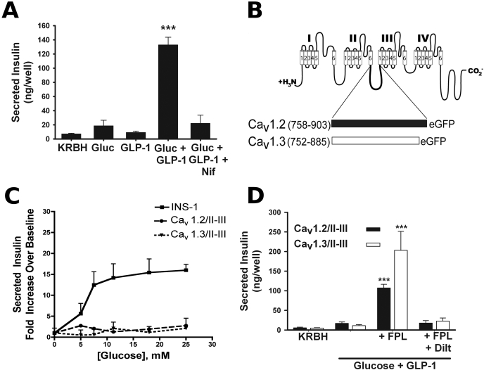
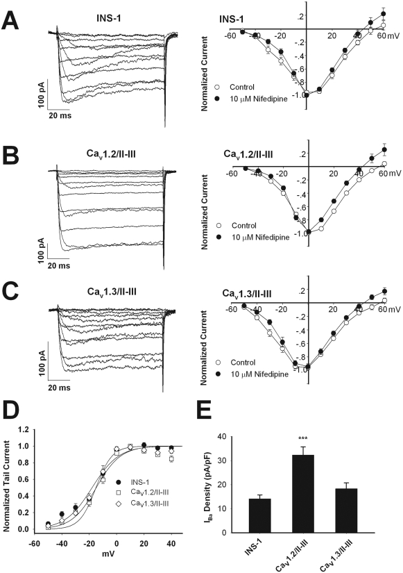
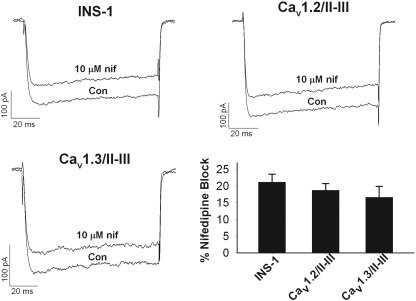
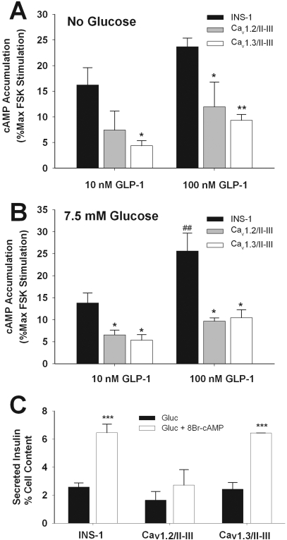
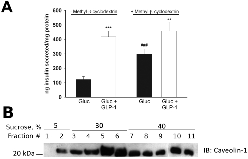
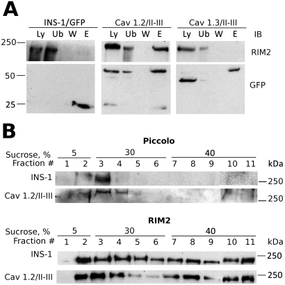
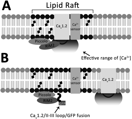
L-type Ca(2+) channels play a key role in the integration of physiological signals regulating insulin secretion that probably requires their localization to specific subdomains of the plasma membrane. We investigated the role of the intracellular II-III loop domains of the L-type channels Ca(v)1.2 and 1.3 in coupling of Ca(2+) influx with glucose-stimulated insulin secretion (GSIS) potentiated by the incretin hormone glucagon-like peptide (GLP)-1. In INS-1 cell lines expressing the Ca(v)1.2/II-III or Ca(v)1.3/II-III peptides, GLP-1 potentiation of GSIS was inhibited markedly, coincident with a decrease in GLP-1-stimulated cAMP accumulation and the redistribution of Ca(v)1.2 and Ca(v)1.3 out of lipid rafts. Neither the Ca(v)1.2/II-III nor the Ca(v)1.3/II-III peptide decreased L-type current density compared with untransfected INS-1 cells. GLP-1 potentiation of GSIS was restored by the L-type channel agonist 2,5-dimethyl-4-[2-(phenylmethyl)benzoyl]-1H-pyrrole-3-carboxylic acid methyl ester (FPL-64176). In contrast, potentiation of GSIS by 8-bromo-cAMP (8-Br-cAMP) was inhibited in Ca(v)1.2/II-III but not Ca(v)1.3/II-III cells. These differences may involve unique protein-protein interactions because the Ca(v)1.2/II-III peptide, but not the Ca(v)1.3/II-III peptide, immunoprecipitates Rab3-interacting molecule (RIM) 2 from INS-1 cell lysates. RIM2, and its binding partner Piccolo, localize to lipid rafts, and they may serve as anchors for Ca(v)1.2 localization to lipid rafts in INS-1 cells. These findings suggest that the II-III interdomain loops of Ca(v)1.2, and possibly Ca(v)1.3, direct these channels to membrane microdomains in which the proteins that mediate potentiation of GSIS by GLP-1 and 8-Br-cAMP assemble.


Figures

Fig. 1.
Fig. 1.
Overexpression of peptides corresponding to the full-length α1 II-III interdomain loops of Cav1.2 and Cav1.3 abrogates potentiation of glucose-stimulated insulin secretion by GLP-1. A, 50 nM GLP-1 robustly potentiates 7.5 mM glucose-stimulated insulin secretion in INS-1 cells. This amplified response is sensitive to the L-VGCC blocker nifedipine (10 μM). ***, P < 0.001 compared with all other conditions (n = 3–10; F = 47.5). B, diagram of the II-III interdomain linker peptide of the L-VGCC subunits Cav1.2 and Cav1.3 fused to GFP that were stably transfected in INS-1 cells. Numbers indicate amino acids included in the fusions from Cav1.2 (M67515.1, GI: 2065770) and Cav1.3 (NM_001128840, GI: 192807299). C, in INS cells, 50 nM GLP-1 amplifies insulin secretion in response to a physiological range of glucose concentrations (5–25 mM). This glucose concentration dependence of insulin release is abolished upon overexpression of the Cav1.2/II-III or the Cav1.3/II-III peptide. Secretion is expressed as -fold increase over KRBH. D, L-type channel “agonist” FPL-64176 (50 μM) rescues GLP-1 potentiation of GSIS in INS-1 cells overexpressing the Cav1.2/II-III or Cav1.3/II-III peptides. This recovery of secretion by FPL-64176 is completely blocked by 500 μM diltiazem. ***, P < 0.001 compared with all other conditions within the same cell line (Cav1.2/II-III: n = 7–13, F = 51.5; and Cav1.3/II-III: n = 5–13, F = 17.4).
Fig. 2.
Fig. 2.
Endogenous voltage-gated Ca2+ channel activity is intact in INS-1 cells overexpressing Cav1.2/II-III or Cav1.3/II-III peptides. A to C, current-voltage relationship curves of IBa measured using whole-cell voltage clamp. Currents were elicited by 100-ms depolarizations from -50 to +60 mV in 10-mV increments, from a holding potential of -70 mV, in the absence (control, ○) or presence of 10 μM nifedipine (•). INS-1 cells (n = 10), Cav1.2/II-III cells (n = 10), and Cav1.3/II-III cells (n = 8). D, peak tail currents recorded upon repolarization to -70 mV after a 100-ms depolarization to the indicated prepulse voltage were normalized and plotted against prepulse voltage. V1/2act values were determined by fitting the data to the equation I/Imax = 1/(1 + exp[-(V - V1/2)/K]), where V is the prepulse potential in millivolts and V1/2 is the prepulse potential at which tail current amplitude is half-maximal. E, whole-cell IBa density (pA/pF) measured at 0 mV from a holding potential of -70 mV. The difference in total whole-cell current density for Cav1.2/II-III cells compared with INS-1 cells and Cav1.3/II-III cells is statistically significant. ***, P < 0.001 (n = 13–15, F = 13.9).
Fig. 3.
Fig. 3.
Overexpression of the Cav1.2/I-III and Cav1.3/II-III loop peptides does not affect the percentage of whole-cell L-type Ca2+ channels in INS-1 cells. Example whole-cell IBa traces were recorded from INS-1, Cav1.2/II-III, and Cav1.3/II-III cells, respectively, showing baseline current (con) and current after coming to equilibrium in the presence of 10 μM nifedipine (nif) applied in the extracellular solution. Cells were held at -70 mV and current elicited by pulsing to +10 mV for 100 ms. Percentage of the whole-cell IBa blocked by 10 μM nifedipine (ns, one-way analysis of variance, F = 0.78) is shown.
Fig. 4.
Fig. 4.
Overexpression of the Cav1.2/II-III or Cav1.3/II-III peptide reduces GLP-1-stimulated cAMP accumulation in INS-1 cells. A, in Cav1.2/II-III or Cav1.3/II-III cells, GLP-1-stimulated cAMP accumulation is reduced compared with control INS-1 cells. Expressed as percentage of cAMP accumulation stimulated with 300 μM forskolin. *, P < 0.05; **, P < 0.01 compared with INS-1 cells (n = 5, F = 5.4). B, addition of glucose (7.5 mM) did not alter cAMP accumulation in INS-1, Cav1.2/II-III, or Cav1.3/II-III cells. *, P < 0.05 compared with INS-1 cells; ##, P < 0.01 compared with 10 nM GLP-1 in INS-1 cells (n = 3, F = 11.5). For all cell lines, the forskolin dose-response curves with or without glucose are not significantly different (see Supplemental Fig. 1). C, overexpression of the Cav1.2/II-III loop, but not the Cav1.3/II-III loop, inhibits potentiation of 7.5 mM GSIS by 1 mM 8-Br-cAMP. Secreted insulin is expressed as percentage of cell content. ***, P < 0.001 relative to glucose alone (n = 3, F = 12.1).
Fig. 5.
Fig. 5.
Displacement of endogenous Cav1.2 or Cav1.3 α1 subunits from detergent-resistant lipid raft membrane fractions by Cav1.2/II-III or Cav1.3/II-III peptides. A, Western blot for Cav1.2 in lysates of INS-1, Cav1.2/II-II, and Cav1.3/II-III cells fractionated over discontinuous sucrose gradients. B, Western blot for Cav1.3 in cell lysates from INS-1, Cav1.2/II-III, and Cav1.3/II-III cells fractionated over discontinuous sucrose gradients. C, Western blot for caveolin-1 in cell lysates from INS-1, Cav1.2/II-III, and Cav1.3/II-III cells fractionated over discontinuous sucrose gradients. Channel antibodies are directed against the II-III interdomain loop of Cav1.2 or Cav1.3. The overexpressed Cav1.2/II-III (46-kDa) or Cav1.3/II-III (43-kDa) peptide was detected in all fractions, with a nonuniform distribution that increased from light to dense fractions (data not shown). Each Western blot is representative of at least three independent experiments.
Fig. 6.
Fig. 6.
β-Methyl cyclodextrin enhances GSIS but does not enhance GLP-1 potentiation of GSIS in INS-1 cells. A, insulin secretion in response to 7.5 mM glucose, but not potentiation of GSIS by 50 nM GLP-1, is enhanced by pretreatment of INS-1 cells with the cholesterol binding agent mβCD (10 mM). ***, P < 0.001; **, P < 0.01 compared with glucose alone; ##, P < 0.01 compared with glucose alone in non-mβCD-treated cell (n = 9, F = 19.9). B, pretreatment of INS-1 cells with 10 mM mβCD does not displace caveolin-1 from the lipid raft fractions of a sucrose density gradient. The Western blot shown is representative of three separate experiments.
Fig. 7.
Fig. 7.
RIM2 preferentially associates with the Cav1.2/II-II peptide and is present in lipid rafts. A, RIM2 coimmunoprecipitates with the II-III loop of Cav1.2. Lysates from INS-1 cells transfected with GFP (INS-1/GFP), Cav1.2/II-III cells, or Cav1.3/II-III cells were incubated with agarose beads fused to a GFP antibody. Samples were analyzed by Western blot. Lane Ly represents 50 μg of protein from cell lysates. Lane Ub is the unbound protein after immunoprecipitation. Lane W represents proteins that were removed through a series of washes. The proteins were eluted from the beads using gentle heating in Laemmli buffer (lane E). For lanes 2 to 4 50 μl of each sample was loaded. Blots were also probed with GFP antibody to confirm the location of the II-III loops. Results shown are representative of three independent experiments. B, Western blot of Piccolo and RIM2 from lysates of INS-1 and Cav1.2/II-III cells fractionated over discontinuous sucrose gradients. Each blot is representative of at least three separate experiments.
Fig. 8.
Fig. 8.
Model for inhibition of GLP-1 potentiation of GSIS by displacement of Cav1.2 or Cav1.3 from lipid rafts. A, intracellular interdomain II-III loops of Cav1.2 and 1.3 direct localization of the respective channels to lipid rafts via binding to raft-resident proteins. In Cav1.2, binding to the raft resident RIM2 anchors the channel in the lipid raft domain. A similar interaction with a yet unidentified protein is proposed to anchor Cav1.3 to lipid rafts. Other proteins required for insulin exocytosis or extracellular signal-regulated kinase 1/2 phosphorylation are also localized to lipid rafts. B, overexpression of the Cav1.2/II-III loop/GFP fusion peptide in INS-1 cells competitively displaces the endogenous Cav1.2 channel from lipid rafts by binding to raft resident protein RIM2, spatially uncoupling them from Ca2+-dependent processes such as GLP-1 potentiation of GSIS. Augmentation of Ca2+ influx via the displaced channels with the L-type channel agonist FPL-64176 can partially compensate for the increased distance between channel pore and Ca2+-sensing proteins involved in secretion (see Fig. 1C).

References

    1. Asfari M, Janjic D, Meda P, Li G, Halban PA, and Wollheim CB (1992) Establishment of 2-mercaptoethanol-dependent differentiated insulin-secreting cell lines. Endocrinology 130 167-178. - PubMed
    1. Balijepalli RC, Foell JD, Hall DD, Hell JW, and Kamp TJ (2006) Localization of cardiac L-type Ca(2+) channels to a caveolar macromolecular signaling complex is required for beta(2)-adrenergic regulation. Proc Natl Acad Sci U S A 103 7500-7505. - PMC - PubMed
    1. Bergsten P (2002) Role of oscillations in membrane potential, cytoplasmic Ca2+, and metabolism for plasma insulin oscillations. Diabetes 51(Suppl 1): S171-S176. - PubMed
    1. Catterall WA (1995) Structure and function of voltage-gated ion channels. Annu Rev Biochem 64 493-531. - PubMed
    1. Cohen AW, Hnasko R, Schubert W, and Lisanti MP (2004) Role of caveolae and caveolins in health and disease. Physiol Rev 84 1341-1379. - PubMed

Publication types

MeSH terms