. 2009 Jun 5;284(23):15951-69.
doi: 10.1074/jbc.M900418200. Epub 2009 Apr 7.

Peroxidase mechanism of lipid-dependent cross-linking of synuclein with cytochrome C: protection against apoptosis versus delayed oxidative stress in Parkinson disease

Affiliations

Peroxidase mechanism of lipid-dependent cross-linking of synuclein with cytochrome C: protection against apoptosis versus delayed oxidative stress in Parkinson disease

Hülya Bayir et al. J Biol Chem. .

Abstract

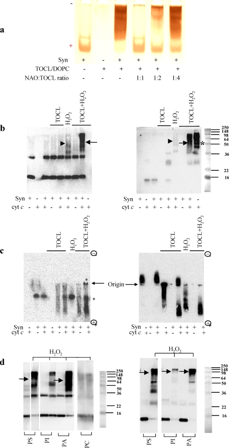
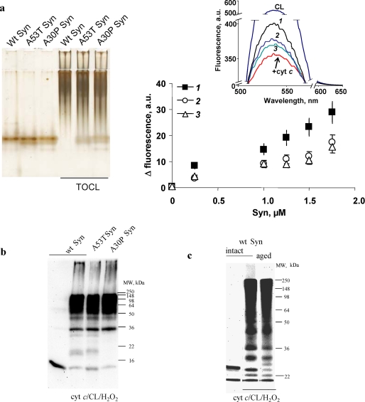
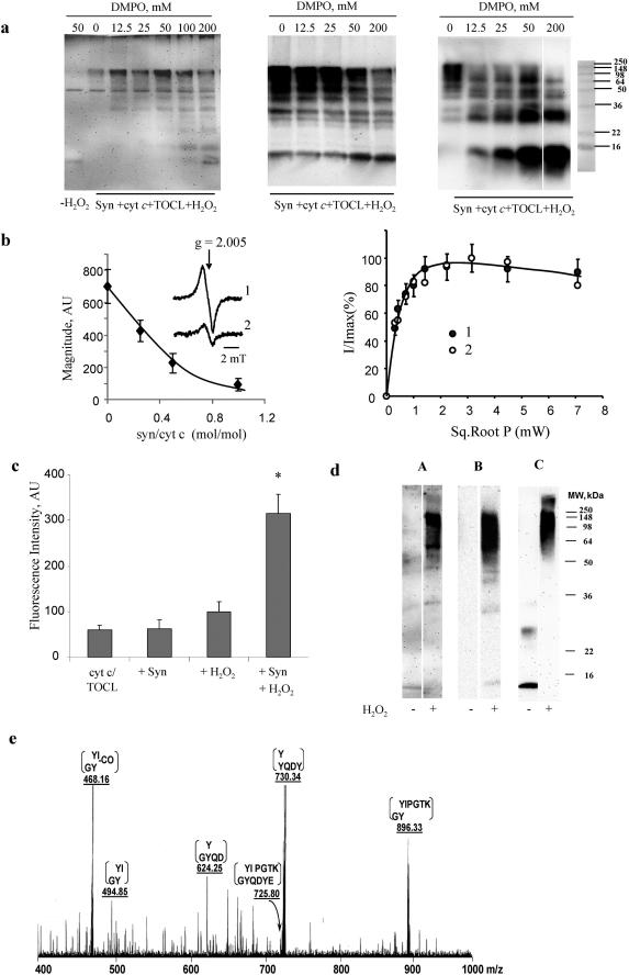
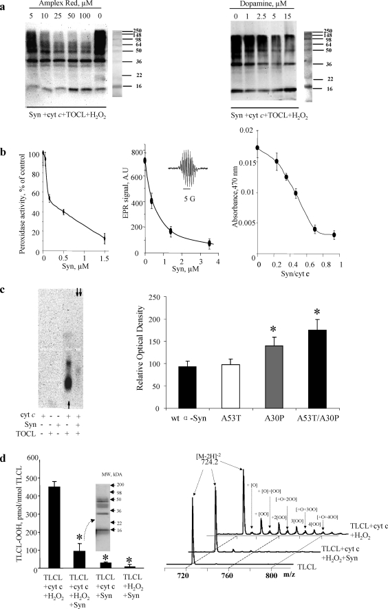
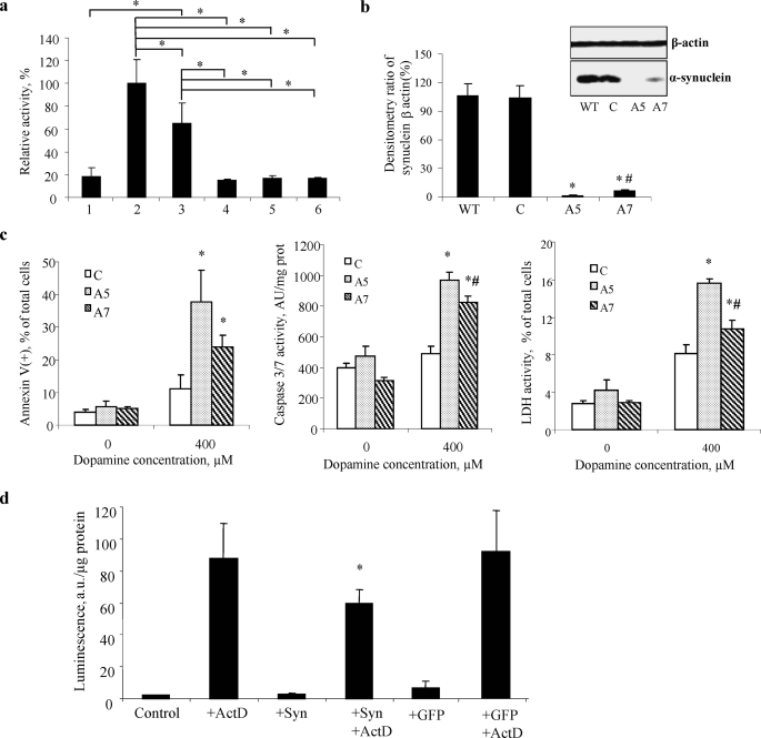
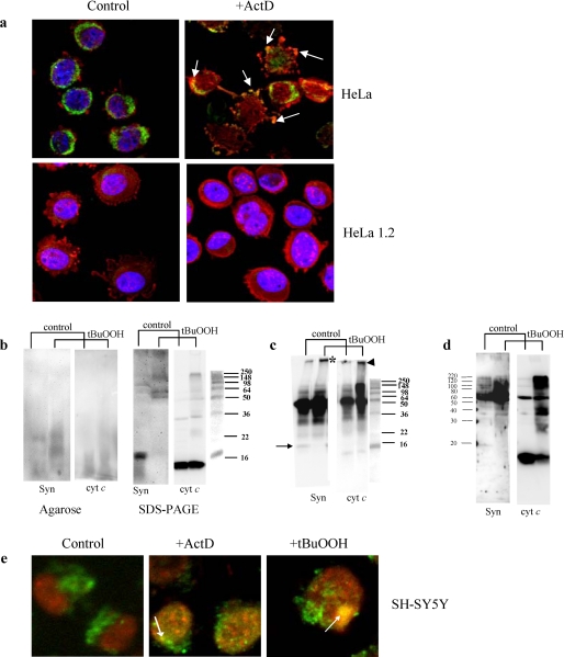
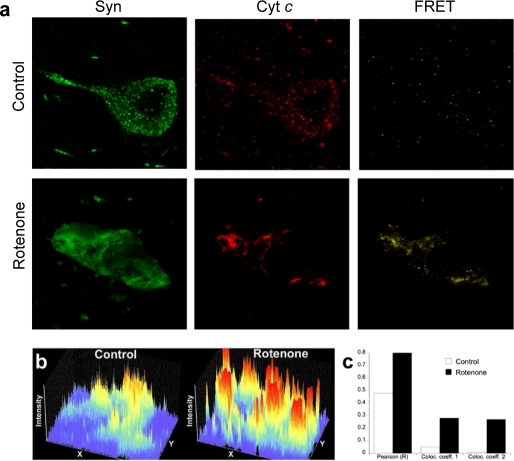
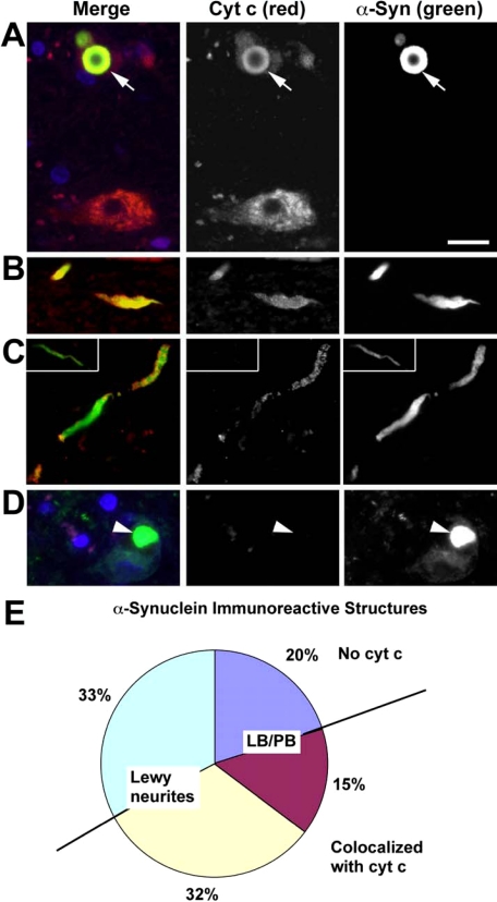
Damage of presynaptic mitochondria could result in release of proapoptotic factors that threaten the integrity of the entire neuron. We discovered that alpha-synuclein (Syn) forms a triple complex with anionic lipids (such as cardiolipin) and cytochrome c, which exerts a peroxidase activity. The latter catalyzes covalent hetero-oligomerization of Syn with cytochrome c into high molecular weight aggregates. Syn is a preferred substrate of this reaction and is oxidized more readily than cardiolipin, dopamine, and other phenolic substrates. Co-localization of Syn with cytochrome c was detected in aggregates formed upon proapoptotic stimulation of SH-SY5Y and HeLa cells and in dopaminergic substantia nigra neurons of rotenone-treated rats. Syn-cardiolipin exerted protection against cytochrome c-induced caspase-3 activation in a cell-free system, particularly in the presence of H(2)O(2). Direct delivery of Syn into mouse embryonic cells conferred resistance to proapoptotic caspase-3 activation. Conversely, small interfering RNA depletion of Syn in HeLa cells made them more sensitive to dopamine-induced apoptosis. In human Parkinson disease substantia nigra neurons, two-thirds of co-localized Syn-cytochrome c complexes occurred in Lewy neurites. Taken together, these results indicate that Syn may prevent execution of apoptosis in neurons through covalent hetero-oligomerization of cytochrome c. This immediate protective function of Syn is associated with the formation of the peroxidase complex representing a source of oxidative stress and postponed damage.


Figures

FIGURE 1.
FIGURE 1.
Electrophoretic evidence for H2O2-induced hetero-oligomerization of Syn with cyt c in the presence of TOCL (a–c) and other anionic phospholipids (d). a, PAGE-based assessment of competitive binding of CL with Syn. A typical polyacrylamide gel was stained for Syn with SilverStain Snap kit. Note that the monomeric form of Syn migrating to the cathode in the absence of TOCL changes its migration behavior upon binding with CL. Titration with different amounts of NAO, which has a known high affinity for CL (16), reconstitutes the Syn migration profile, because of competitive binding of TOCL with NAO. The amount of free Syn was quantified by densitometry using Bio-Rad Multi-Imager and Multi-Analyst software. b, SDS-PAGE of aggregates formed after incubation of Syn with cyt c/CL/H2O2. Staining with anti-Syn (left panel) and anti-cyt c antibodies (right panel) reveals that all for components Syn, cyt c, TOCL, and H2O2 were required for the formation of hetero-oligomers (containing both Syn and cyt c, detectable on both panels, arrows). A weak H2O2-induced aggregation of Syn with cyt c was observed in the absence of TOCL (arrowheads) in line with the known induction of peroxidase activity of cyt c during incubation with H2O2 (8). As expected, significant aggregation of cyt c occurs (asterisk) in the absence of Syn as cyt c-TOCL complexes exert significant peroxidase activity causing H2O2-dependent aggregation (12). However, the aggregates are formed due to homo-oligomerization of cyt c, and they do not contain Syn (compare with the lack of staining for Syn on the left panel). c, native electrophoresis in agarose gel and Western blotting of complexes formed after incubation of Syn with cyt c and TOCL. Staining was with anti-Syn (left panel) and anti-cyt c antibodies (right panel). Note that Syn and cyt c did not interact with each other in the absence of TOCL. In the absence of H2O2, Syn and cyt c migrated mostly as single monomeric proteins. TOCL changed migration profile of Syn and cyt c. TOCL plus H2O2 caused oligomerization of Syn-cyt (asterisks) as evidenced by the appearance of aggregates at the origin of the gel and dense smeared pattern on the bottom of the gel. Thus, all four components, Syn, cyt c, CL, and H2O2, were required for the catalytic formation of cross-linked polymers. Note that no nonspecific reactivity was displayed by anti-Syn or anti-cyt c antibodies to either monomeric or aggregated forms of cyt c and Syn, respectively. d, SDS electrophoresis of aggregates formed after incubation of Syn with cyt c and H2O2 in the presence of different anionic phospholipids as follows: PS, PI, and PA. A nonanionic phospholipid PC was used as a control (shown for Syn). Staining with anti-Syn antibody (left panel) and anti-cyt c antibody (right panel) revealed that all four components, Syn, cyt c, H2O2, and anionic phospholipids, were required (arrows) for the formation of hetero-oligomers (containing both Syn and cyt c and detectable on both panels). Note that all four tested phospholipids were active in inducing H2O2-dependent hetero-oligomerization of Syn with cyt c; PI was significantly less effective than other phospholipids. In the absence of cyt c, none of the phospholipids tested caused oligomerization of Syn (data not shown). Typical gels representative of five experiments are shown.
FIGURE 2.
FIGURE 2.
Formation of hetero-oligomeric complexes and covalent cross-linked aggregates of cyt c/TOCL with wild-type Syn (intact and aged) and its mutants A53T and A30P. a, native PAGE of Syn-cyt c-TOCL hetero-oligomeric complexes (staining with SilverSNAP stain kit, ThermoFisher) (left panel). Syn (8 μm) was incubated with TOCL/DOPC liposomes (TOCL/Syn ratio 20:1) and cyt c (8 μm) for 60 min at 37 °C. Assessment of Syn binding to CL/cyt c using NBD-CL fluorescence (right panel) is shown. Inset, typical fluorescence spectra obtained from the following: CL/NBD-CL liposomes (uppermost curve), CL/NBD-CL-cyt c complexes (lowest curve), and CL/NBD-CL/cyt c after titration with wild-type (Wt) Syn (1) and mutant forms of Syn A53T (2) and A30P (3) (1.75 μm). b, SDS-PAGE (12%) with subsequent Western blot analysis (using anti-Syn antibody) of cross-links formed after incubation of wild-type (wt) Syn, A53T Syn, and A30P Syn with cyt c/TOCL in the presence of H2O2. Wild-type Syn, A30P, and A53T mutants underwent hetero-oligomeric covalent cross-linking with cyt c in a similar way. c, SDS-PAGE (7.5%) with subsequent Western blot analysis (using anti-Syn antibody) of Syn-cyt c-CL covalent cross-links formed by incubation of nonfibrillated and fibrillated (aged) Syn with cyt c/TOCL in the presence of H2O2. Comparable covalent cross-linking of nonfibrillated and fibrillated Syn was observed in the presence of cyt c, TOCL, and H2O2.
FIGURE 3.
FIGURE 3.
Evidence for the involvement of peroxidase reactive intermediates in hetero-oligomerization of Syn-cyt c-TOCL in the presence of H2O2. a, immuno-spin trapping of protein-derived radicals during H2O2-dependent hetero-oligomerization of Syn with cyt c in the presence of TOCL. Staining of polyacrylamide gels with anti-DMPO antibody (left panel) demonstrates the production of protein-derived radicals. Staining with anti-Syn (middle panel) and anti-cyt c (right panel) antibodies is shown. Note that the production of hetero-oligomeric forms of Syn-cyt c-TOCL aggregates is progressively inhibited by increasing concentrations of DMPO indicating that the formation of DMPO adducts competitively inhibited oligomerization. No oligomerization of Syn with cyt c and TOCL occurred in the absence of H2O2 (data not shown). Typical gels representative of three experiments are shown. b, low temperature EPR measurements of H2O2-induced protein-derived (tyrosyl) radicals of cyt c-CL complexes are shown in the left panel. Syn quenched the formation of tyrosyl radicals of cyt c-CL complexes in a concentration-dependent manner. The inset shows a typical EPR spectra of protein-derived (tyrosyl) radicals of cyt c-CL complexes in the absence (trace 1) and presence of Syn (trace 2), cyt c/α-synuclein = 1:0.5. Right panel shows power-saturation curves of protein-derived tyrosyl radical EPR signals of cyt c-CL complexes (trace 1) and cyt c-CL-Syn complexes (trace 2) (cyt c/α-synuclein = 1:0.5). c, assessments of dityrosine cross-links formed by incubation of Syn-cyt c-TOCL with H2O2 by fluorescence intensity (λex 315 nm and λem 420 nm). Formation of dityrosine adducts increased by 1.6-fold upon addition of H2O2 to cyt c/TOCL. When Syn was present in this incubation system along with H2O2, the fluorescence intensity was further increased by 3.2-fold (mean ± S.D., *, p < 0.05 versus cyt c/TOCL/H2O2). d, SDS-PAGE with subsequent Western blot analysis of covalent conjugates with dityrosine cross-links formed from syn-cyt c-TOCL hetero-oligomeric complexes in the presence of H2O2. Staining was performed using anti-dityrosine antibody (lanes A), anti-Syn antibody (lanes B), and anti-cyt c antibody (lanes C). A strong positive signal was observed with anti-dityrosine antibody in cross-linked high molecular weight aggregates (nondissociable in SDS). e, MS/MS spectrum of m/z 725.8 (M + 2H)2+ species. This species was obtained from the digestion of high molecular weight aggregates of Syn-cyt c-CL formed in the presence of H2O2. Note the presence of ions at m/z 468, 495, 625, 730, and 896 corresponding to various daughter ions of the Syn-(132–137) and cyt c-(74–79) cross-link.
FIGURE 4.
FIGURE 4.
Syn is preferred substrate for reactive intermediates of the peroxidase complex. a, effects of two prototypical peroxidase phenolic substrates Amplex Red (left panel) and DA (right panel) on oligomerization of Syn-cyt c-TOCL in the presence of H2O2. Staining with anti-Syn antibody. Note that both compounds caused concentration-dependent inhibition of protein hetero-oligomerization. Typical gels representative of three experiments are shown. b, inhibition of peroxidase activity of Syn-cyt c-TOCL complexes with Amplex Red, DA, and etoposide. Left panel, Syn inhibits the peroxidase activity of Syn-cyt c-TOCL complexes, with Amplex Red as a substrate, in a concentration-dependent manner. Note that the IC50 for Syn was 0.2 μm in the presence of 50 μm Amplex Red. The results of three independent experiments are shown. Middle panel, Syn inhibits the peroxidase activity of Syn-cyt c-TOCL complexes, with etoposide as a substrate, in a concentration-dependent manner. Note that the IC50 for Syn was 0.5 μm in the presence of 100 μm etoposide. A typical ESR spectrum of etoposide phenoxyl radical generated by the H2O2-dependent peroxidase activity is shown (inset). Right panel, Syn inhibits the peroxidase activity of Syn-cyt c-TOCL complexes, with DA as a substrate (200 μm). Data are presented as mean ± S.D. (n = 3). c, native agarose gels of Syn-cyt c-CL and cyt c-CL complexes stained for peroxidase activity with SuperSignal West Pico chemiluminescence substrate (left panel). Note that the peroxidase activity of cyt c-TOCL complexes (single arrow) toward the chemiluminescence substrate is inhibited when Syn is included in the preformed complex (double arrows) and then exposed to H2O2/chemiluminescence substrate. Typical gels representative of three experiments are shown. Quantification of residual peroxidase activity of wild-type (wt) and mutated forms of Syn (A53T, A30P, A53T/A30P)-cyt c-TOCL complexes (right panel). The peroxidase activity of cyt c-TOCL complexes was higher in the presence of Syn mutants (A30P and A53T/A30P) compared with wild-type Syn (mean ± S.D.; *, p < 0.05 versus wild-type Syn). d, Syn protects polyunsaturated TLCL against oxidation induced by cyt c/H2O2. Cyt c-induced accumulation of oxidized TLCL (as assessed by fluorescence HPLC (left panel) and MS analysis of doubly charged ions [M − 2H]2− (right panel)) was markedly suppressed by Syn. This was accompanied by the formation of high molecular weight aggregates of Syn-cyt c-TLCL (see Western blot with Syn antibody (left panel, inset). Data are presented as mean ± S.E.; *, p < 0.05 versus TLCL + cyt c + H2O2. Accumulation of monohydroperoxy/monohydroxy derivatives of TLCL molecular species was observed after incubation of liposomes with cyt c and H2O2 (m/z 724.2 + [O] = 732.2; 724.2 + [OO] = 739.7; 724.2 + [O] + [OO] = 748.2; 724.2 + [2OO] = 755.9; 724.2 + [O] + [2OO] = 763.8; 724.2 + [3OO] = 771.8; 724.2 + [O] + [3OO] = 779.7; 724.2 + [4OO] = 787.9; and 724 + [O] + [4OO] = 795.7). This effect was markedly reduced by Syn. Representative mass spectra from three independent experiments are presented.
FIGURE 5.
FIGURE 5.
Syn and Syn-CL complex inhibits apoptotic caspase activation. a, Syn-TOCL complexes inhibit caspase-3 activated by cyt c/dATP in a cell-free proapoptotic system (S-100 fraction isolated from HL-60 cells). Note that Syn in combination with TOCL was most effective in preventing cyt c-dependent caspase-3 activation. Wild-type Syn and two mutants, A53T and A30P, displayed equal inhibitory effects against caspase-3 activation after preincubation with cyt c/TOCL/H2O2. S-100 (5 μg of protein/μl) fractions were incubated with 1 μm cyt c for 90 min at 37 °C, in the absence and in the presence of complex of Syn-TOCL. Bar 1, S-100; bar 2, 1 + cyt c; bar 3, 2 + TOCL/Syn; bar 4, complex of wild-type Syn-TOCL-cyt c was preincubated with H2O2 and then added into 1; bar 5, complex of A53T Syn-TOCL-cyt c was preincubated with H2O2 and then added into 1; bar 6, complex of A30P Syn-TOCL-cyt c was preincubated with H2O2 and then added into 1. All the data are presented as % of caspase-3 activation in complete system (bar 2) taken as 100%. Data are presented are means ± S.D. (n = 6). The statistical significance was calculated using analysis of variance, followed by Tukey's procedure with the family-wise error rate of p < 0.05 to perform our pairwise comparisons of selected group means. Asterisk indicates a statistically significant difference resulting from the Tukey comparisons. b, densitometric and Western blot analysis (inset) with anti-Syn antibody in wild-type (WT), nontargeting siRNA (C), two different clones (A5 and A7) of HeLa Syn knockdown. Data are presented as mean ± S.D. (n = 3/condition). *, p < 0.05 versus wild type; #, p < 0.05, A7 versus A5. c, biomarkers of apoptosis-PS externalization (left panel) and caspase-3/7 activation (middle panel) increase and cell viability (right panel) decrease in Syn-deficient HeLa A5 and A7 cells after exposure to DA. Data are presented as mean ± S.D. (n = 3/condition). *, p < 0.05, 400 μm versus 0 μm; #, p < 0.05, A7 versus A5. d, delivery of Syn into MECs confers resistance to proapoptotic caspase-3/7 activation. Note that robust activation of caspase-3/7 MECs stimulated with ActD was markedly attenuated in Syn-containing MECs (delivered by the direct introduction of Syn into cells). Delivery of a nonspecific protein, green fluorescent protein (GFP), exerted no effect on ActD-induced caspase-3/7 activation in MECs. Data are presented as mean ± S.D. (n = 4/condition). *, p < 0.05, ActD versus ActD + Syn.
FIGURE 6.
FIGURE 6.
Detection of inclusions and co-localization of hetero-polymerized Syn and cyt c aggregates in HeLa and SH-SY5Y cells after exposure to ActD or t-BuOOH. a, immunostaining of cyt c (green) and Syn (red) in intact HeLa cells and cyt c-deficient HeLa 1.2 cells. In control cells, cyt c appears in a punctate, perinuclear pattern of staining with little overlap with cytosolic Syn staining. ActD induces cytoplasmic blebbing, nuclear fragmentation, and transition from perinuclear to a diffuse staining pattern for cyt c. Focal regions of accentuated Syn staining, suggestive of aggregation, are observed, and this frequently co-localizes with focal regions of accentuated cyt c staining in cells with both preserved and fragmented nuclear contours. HeLa 1.2 clones engineered to express decreased levels of cyt c show a similar distribution of cyt c and Syn, although there is less cyt c immunoreactivity. HeLa 1.2 cells show preserved perinuclear cyt c and reduced propensity for clumped or aggregated Syn in response to the same doses of ActD, and there is no evidence of Syn and cyt c co-localization in these cells. b, Western blot analysis of Syn-cyt c aggregates after native agarose gel electrophoresis (left panel) and SDS-PAGE (right panel) in HeLa cells exposed to t-BuOOH. Left panel, staining with anti-Syn (Syn) and anti-cyt c antibodies (cyt c) reveals the appearance of co-localized hetero-oligomers after treatment of cells with t-BuOOH. Right panel, staining with anti-Syn (Syn) and anti-cyt c antibodies (cyt c) demonstrates disappearance of monomeric form of Syn, and accumulation of very high molecular weight Syn aggregates partially overlapping with aggregated forms of cyt c. Typical gels representative of three experiments are shown. c, detection of Syn-cyt c hetero-oligomers in HeLa cells exposed to t-BuOOH using immunoprecipitation with anti-Syn antibodies and Western blot analysis with anti-Syn and anti-cyt c antibodies after SDS-PAGE. Note that proteins immunoprecipitated with anti-Syn antibody contained high molecular weight aggregates positive for both Syn (asterisk) and cyt c (arrowhead) after treatment with t-BuOOH. Bands around 50 kDa likely correspond to heavy chains of immunoglobulins remaining after immunoprecipitation. Arrow indicates Syn monomer. Typical gels representative of three experiments are shown. d, Western blot analysis of Syn-cyt c aggregates after SDS-PAGE in mitochondria t-BuOOH from HeLa cells pretreated with t-BuOOH. Staining with anti-Syn (Syn) and anti-cyt c antibodies (cyt c) demonstrates accumulation of very high molecular weight Syn aggregates partially overlapping with aggregated forms of cyt c. Note that mitochondria from control cells contain significant amounts of monomeric cyt c but hardly detectable levels of monomeric Syn. After treatment with t-BuOOH, polymerized forms of Syn and cyt c are readily detectable, and the monomeric form of cyt c is decreased. Typical gels representative of three experiments are shown. e, immunostaining of cyt c (green) and Syn (red) in SH-SY5Y cells. Note that both proapoptotic agents, t-BuOOH and ActD, caused the appearance of LB-like inclusions revealed as yellow color in merged images (arrows). In normal SH-SY5Y cells, cyt c had a punctate mitochondrial distribution, and Syn was localized in the cytoplasm and perinuclear area. Following ActD or t-BuOOH treatment, Syn displayed aggregated pattern of distribution and overlapped with anti-cyt c staining (yellow spots on merged images).
FIGURE 7.
FIGURE 7.
Syn distribution and co-localization with cyt c in substantia nigra dopaminergic neurons after rotenone. a, brain sections through substantia nigra were double-labeled for cyt c (red) and Syn (green) and imaged by laser scanning confocal microscopy. Note the increased fluorescence and altered distribution of Syn after rotenone. Additionally, there is FRET between cyt c and Syn, strongly suggesting direct protein/protein interactions. b, fluorescence intensity profiles of Syn immunoreactivity in single dopaminergic neuron from control and rotenone-treated rats. Each fluorescence intensity profile is from an identical rectangular region of interest encompassing part of a single dopaminergic neuron. After rotenone, the neuronal levels of Syn increase, and its distribution changes from relatively diffuse to markedly punctate. c, quantification of the co-localization of Syn with cyt c in substantia nigra from a control animal and a rotenone-treated animal. After rotenone treatment there is marked increase in the Pearson correlation coefficient (R) and in the co-localization coefficients for both channel 1 (cyt c) and channel 2 (Syn). Data are from confocal images (×60) that each contain ∼20 dopaminergic neurons and surrounding neuropil.
FIGURE 8.
FIGURE 8.
Co-localization of Syn and cyt c in human PD/Lewy body disease substantia nigra. Midbrain sections from patients with PD/Lewy body disease were immunolabeled for cyt c (red) and Syn (green). 4′,6-Diamidino-2-phenylindole (blue) was used to stain nuclei. Normal nigral neurons exhibit a granular cytoplasmic staining pattern for cyt c, consistent with its mitochondrial localization (A, lower neuron). In contrast, the neuron containing a LB (arrow) shows light, diffuse cytoplasmic cyt c staining and co-localization of cyt c and Syn (yellow) in the LB (A, upper neuron). Many Lewy neurites also showed co-localization of cyt c and Syn (B and C), although cyt c negative Lewy neurites (asterisk) were also observed (inset, C). Note the absence of red staining in the LB containing neuron (arrowhead) when nonimmune rabbit antiserum was substituted for antibody to cyt c as a negative staining control (D). More than 1,200 consecutive Syn immunoreactive structures from four cases were scored by location in the neuronal soma (Lewy body/pale body, LB/PB) or in neuritic processes (Lewy neurites). These were analyzed for presence or absence of cyt c co-localization. The pie chart (E) shows Lewy neurites without cyt c (light green-blue), LB/PB without cyt c (dark blue), LB/PB co-localizing with cyt c (magenta), and Lewy neurites co-localizing with cyt c (yellow). Note that nearly half of all Syn immunoreactive structures show co-localization with cyt c (yellow and red combined). Moreover, more than two-thirds of Syn-cyt c co-localizing structures involved neurites (yellow).

References

    1. Betarbet R., Sherer T. B., Di Monte D. A., Greenamyre J. T. ( 2002) Brain Pathol. 12, 499– 510 - PMC - PubMed
    1. Irizarry M. C., Growdon W., Gomez-Isla T., Newell K., George J. M., Clayton D. F., Hyman B. T. ( 1998) J. Neuropathol. Exp. Neurol. 57, 334– 337 - PubMed
    1. Baba M., Nakajo S., Tu P. H., Tomita T., Nakaya K., Lee V. M., Trojanowski J. Q., Iwatsubo T. ( 1998) Am. J. Pathol. 152, 879– 884 - PMC - PubMed
    1. Dickson D. W. ( 2001) Curr. Opin. Neurol. 14, 423– 432 - PubMed
    1. Shults C. W. ( 2006) Proc. Natl. Acad. Sci. U. S. A. 103, 1661– 1668 - PMC - PubMed

Publication types

MeSH terms