. 2009 Apr 21;106(16):6706-11.
doi: 10.1073/pnas.0810599106. Epub 2009 Apr 7.

A point mutation in TRPC3 causes abnormal Purkinje cell development and cerebellar ataxia in moonwalker mice

Affiliations

A point mutation in TRPC3 causes abnormal Purkinje cell development and cerebellar ataxia in moonwalker mice

Esther B E Becker et al. Proc Natl Acad Sci U S A. .

Abstract

The hereditary ataxias are a complex group of neurological disorders characterized by the degeneration of the cerebellum and its associated connections. The molecular mechanisms that trigger the loss of Purkinje cells in this group of diseases remain incompletely understood. Here, we report a previously undescribed dominant mouse model of cerebellar ataxia, moonwalker (Mwk), that displays motor and coordination defects and loss of cerebellar Purkinje cells. Mwk mice harbor a gain-of-function mutation (T635A) in the Trpc3 gene encoding the nonselective transient receptor potential cation channel, type C3 (TRPC3), resulting in altered TRPC3 channel gating. TRPC3 is highly expressed in Purkinje cells during the phase of dendritogenesis. Interestingly, growth and differentiation of Purkinje cell dendritic arbors are profoundly impaired in Mwk mice. Our findings define a previously unknown role for TRPC3 in both dendritic development and survival of Purkinje cells, and provide a unique mechanism underlying cerebellar ataxia.


Conflict of interest statement

The authors declare no conflict of interest.

Figures
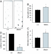
Fig. 1.
Fig. 1.
Ataxic behavior in Mwk/+ mice. (A) Representative footprint patterns of wild-type and mutant mice at 2 months. Footprint patterns were quantitatively assessed for gait width (B) and step alternation uniformity (C). Mwk/+ mice displayed significantly wider gait (33 ± 2 mm vs. 27 ± 1 mm) and irregular step alternation (alternation coefficient 0.36 ± 0.035 vs. 0.24 ± 0.02) compared with wild-type controls (mean ± SEM, n = 8, P < 0.05, ANOVA followed by Fisher's PLSD post hoc test). (D) Static rod performance of wild-type and mutant mice at 2 months. Mwk/+ mice were significantly impaired in their ability to walk across the beam (35.7 ± 13.25 sec vs. 180 ± 0 sec; mean ± SEM, n = 8, P < 0.0001, ANOVA followed by Fisher's PLSD post hoc test).
Fig. 2.
Fig. 2.
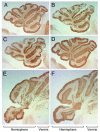
Progressive Purkinje neuron loss in the Mwk/+ cerebellum. Calbindin-stained parasagittal sections from 6-month-old (A), 9-month-old (B), 12-month-old Mwk/+ (C), and wild-type mice (D). Coronal sections of 6-month-old Mwk/+ (E) and wild-type (F) mice show pronounced loss of Purkinje cells in the lateral cerebellar hemispheres of mutant mice.
Fig. 3.
Fig. 3.
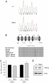
The Mwk mutation is identified in the Trpc3 gene. (A) Sequence analysis of the Trpc3 locus in wild-type and Mwk/+ DNA. An A1903G transversion was detected in Mwk/+ mice but was not present in the parental substrains. (B) Schematic of the 6-transmembrane TRPC3 channel protein. The expanded S4/S5 linker contains the Mwk mutation (T635A). Multiple amino acid sequence alignment of this region between orthologous TRPC3 and related (TRPC6, TRPC7) proteins from different mammalian species shows a very high degree of conservation. (C) TRPC3 and TRPC1 RNA levels normalized against calbindin RNA levels in cerebella from 3-week-old wild-type and Mwk/+ mice. (D) TRPC3 and actin protein levels in cerebellar lysates from 3-week-old wild-type and Mwk/+ mice.
Fig. 4.
Fig. 4.
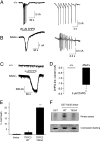
The Mwk mutation results in altered TRPC3 phosphorylation and gating that promotes aberrant channel opening and cell death. Inward current in wild-type (A) and Mwk/+ (B) slices upon application of 20 μM DHPG. The spikes recorded in Mwk/+ Purkinje cells were significantly smaller and occurred at a faster interspike interval (Mwk/+: 0.14 ± 0.02 s interspike interval, 1.156 ± 0.307 nA spike amplitude, n = 5; wild-type: 1.36 + 0.48 s interspike interval, 4.188 + 0.1 nA spike amplitude, n = 3; mean ± SEM, P < 0.05, unpaired Student's t test). Representative examples of Purkinje cells are shown. Spikes are expanded in right panels. (C) Upon application of 5 μM DHPG, there was hardly any inward current in wild-type cells (Upper), but a clear inward current in Mwk/+ Purkinje cells (Lower). (D) Summary of 5-μM DHPG-evoked currents (Mwk/+: 1.635 ± 0.393 pA, n = 6; wild-type: 0.044 ± 0.03 pA, n = 5; mean ± SEM, P < 0.01, unpaired Student's t test). (E) Transient overexpression of Mwk TRPC3 (T635A) but not wild-type TRPC3 significantly induced cell death in NSC-34 cells (25 ± 0.577 vs. 2 ± 1.528, n = 3; mean ± SEM, P < 0.0001, ANOVA followed by Fisher's PLSD post hoc test). (F) In vitro kinase assays using PKCγ with recombinant GST, wild-type TRPC3 GST-S4/S5 linker, and Mwk TRPC3 GST-S4/S5 linker (T635A).
Fig. 5.
Fig. 5.
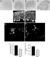
TRPC3 is a critical regulator of dendritic growth and differentiation in cerebellar Purkinje cells. (A–D) In situ hybridization images for Trpc3 in wild-type mouse cerebellum at 8 days (A), 13 days (B), 18 days (C), and 56 days (D) of age. (E and F) Cerebellar sections of 3-week-old wild-type (E) and Mwk/+ (F) mice subjected to immunohistochemistry using an anti-calbindin antibody. (G–J) Organotypic slice cultures (P8 + 12) prepared from wild-type mice (G) and Mwk/+ (H) littermates were stained with an anti-calbindin antibody. Arrowheads indicate dendritic trees. Mwk/+ Purkinje cells had a significantly shorter longest dendrite (I) (44.8 ± 2.5 μm vs. 74.18 ± 3.6 μm) and significantly reduced areas of the dendritic arborization (J) (1358.7 ± 119.9 μm2 vs. 3487.9 ± 225.14 μm2). Results shown are representative of 3 independent experiments (mean ± SEM, n = 59, P < 0.0001, ANOVA followed by Fisher's PLSD post hoc test).

References

    1. Duenas AM, Goold R, Giunti P. Molecular pathogenesis of spinocerebellar ataxias. Brain. 2006;129:1357–1370. - PubMed
    1. Klockgether T. Recent advances in degenerative ataxias. Curr Opin Neurol. 2000;13:451–455. - PubMed
    1. Taroni F, DiDonato S. Pathways to motor incoordination: The inherited ataxias. Nat Rev Neurosci. 2004;5:641–655. - PubMed
    1. Matilla-Duenas A. The highly heterogeneous spinocerebellar ataxias: From genes to targets for therapeutic intervention. Cerebellum. 2008;7:97–100. - PubMed
    1. Lim J, et al. A protein-protein interaction network for human inherited ataxias and disorders of Purkinje cell degeneration. Cell. 2006;125:801–814. - PubMed

Publication types