Review
. 2009 Apr;6(4):191-203.
doi: 10.1038/nrurol.2009.27.

Imaging techniques for prostate cancer: implications for focal therapy

Affiliations
Review

Imaging techniques for prostate cancer: implications for focal therapy

Baris Turkbey et al. Nat Rev Urol. 2009 Apr.

Abstract

The multifocal nature of prostate cancer has necessitated whole-gland therapy in the past; however, since the widespread use of PSA screening, patients frequently present with less-advanced disease. Many men with localized disease wish to avoid the adverse effects of whole-gland therapy; therefore, focal therapy for prostate cancer is being considered as a treatment option. For focal treatment to be viable, accurate imaging is required for diagnosis, staging, and monitoring of treatment. Developments in MRI and PET have brought more attention to prostate imaging and the possibility of improving the accuracy of focal therapy. In this Review, we discuss the advantages and disadvantages of conventional methods for imaging the prostate, new developments for targeted imaging, and the possible role of image-guided biopsy and therapy for localized prostate cancer.


Conflict of interest statement

Competing interests

The authors and the Journal Editor A Hay declared no competing interests. The CME questions author CP Vega declared that he has served as an advisor or consultant to Novartis, Inc.

Figures

Figure 1
Figure 1
MRI of an 82-year-old man with prostate cancer and extracapsular extension. a | The axial T2-weighted image demonstrates a lesion (arrow) in the left mid anterior peripheral zone, with frank bulging secondary to extracapsular extension. b | The coronal T2-weighted image also shows the extracapsular extension (arrow). c | The lesion (arrow) is positive on an apparent diffusion coefficient map of diffusion-weighted MRI.
Figure 2
Figure 2
MRI of a 53-year-old man with prostate cancer. a | The axial T2-weighted image demonstrates a midline low-signal-intensity focus at the mid peripheral zone (arrow), indicating the prostate tumor. b | The sagittal T2-weighted image also demonstrates the midline low-signal-intensity focus (arrow). c | The lesion is positive on an apparent diffusion coefficient map (arrows). d | On the two-compartment pharmacokinetic model analysis of dynamic contrast-enhanced MRI, the Ktrans and kep values of the tumor are high, which indicates a high likelihood of malignancy. e | On magnetic resonance spectroscopy, the choline-to-citrate ratio is increased, indicating malignancy.
Figure 3
Figure 3
Work flow of MRI–TRUS fusion-guided prostate biopsy. MRI is performed before the biopsy. The images are segmented, and the target tumor is identified. The patient then undergoes 3D ultrasound acquisition, and the MRI and TRUS images are combined (registration). By use of real-time TRUS, and the corresponding MRI, the biopsy is targeted towards the tumor. Abbreviations: 3D, three-dimensional; DCE, dynamic contrast-enhanced MRI; DWI, diffusion-weighted MRI; MRS, magnetic resonance spectroscopy; T2W, T2-weighted MRI; TRUS, transrectal ultrasonography. Permission obtained from Sheng Xu and Jochen Kruecker, Philips Research North America, Briarcliff Manor, NY, 10510, USA.
Figure 4
Figure 4
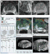
MRI of a 69-year-old man without previous documentation of prostate cancer. a | The axial T2-weighted image demonstrates a round-shaped, low-signal-intensity lesion at the right mid peripheral zone, which is suspicious for cancer (arrow). b | The sagittal T2-weighted image also shows the low-signal-intensity lesion (arrow). c | The apparent diffusion coefficient map of diffusion-weighted MRI is positive (arrow). d | The top two images are screen shots of the fused MRI–TRUS image guidance, illustrating the T2-weighted MRI fused with reference TRUS volume (color map). The bottom two images show the real-time TRUS and target information, corresponding to real-time TRUS at the time of needle deployment. The patient underwent a fused MRI–TRUS-guided biopsy, which was Gleason score 3 + 4 in 70% of the specimen. Permission obtained from Sheng Xu and Jochen Kruecker, Philips Research North America, Briarcliff Manor, NY 10510, USA. Abbreviation: TRUS, transrectal ultrasonography.

References

    1. American Cancer Society (online 2008) Cancer Facts & Figures 2008. 2009 http://www.cancer.org/downloads/STT/2008CAFFfinalsecured.pdf.
    1. Hricak H, Choyke PL, Eberhardt SC, Leibel SA, Scardino PT. Imaging prostate cancer: a multidisciplinary perspective. Radiology. 2007;243:2028–2053. - PubMed
    1. Cornud F, et al. Endorectal color Doppler sonography and endorectal MR imaging features of nonpalpable prostate cancer: correlation with radical prostatectomy findings. AJR Am J Roentgenol. 2000;175:1161–1168. - PubMed
    1. Yang JC, et al. Contrast-enhanced gray-scale transrectal ultrasound-guided prostate biopsy in men with elevated serum prostate-specific antigen levels. Acad Radiol. 2008;15:1291–1297. - PubMed
    1. Tang J, Yang JC, Li Y, Li J, Shi H. Peripheral zone hypoechoic lesions of the prostate: evaluation with contrast-enhanced gray scale transrectal ultrasonography. J Ultrasound Med. 2007;26:1671–1679. - PubMed

MeSH terms