. 2009;4(4):e5136.
doi: 10.1371/journal.pone.0005136. Epub 2009 Apr 8.

The anti-apoptotic activity of BAG3 is restricted by caspases and the proteasome

Affiliations

The anti-apoptotic activity of BAG3 is restricted by caspases and the proteasome

Victoria M Virador et al. PLoS One. 2009.

Abstract

Background: Caspase-mediated cleavage and proteasomal degradation of ubiquitinated proteins are two independent mechanisms for the regulation of protein stability and cellular function. We previously reported BAG3 overexpression protected ubiquitinated clients, such as AKT, from proteasomal degradation and conferred cytoprotection against heat shock. We hypothesized that the BAG3 protein is regulated by proteolysis.

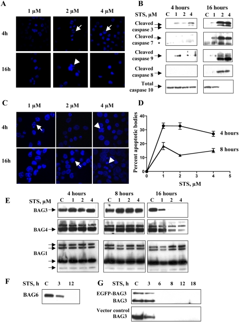
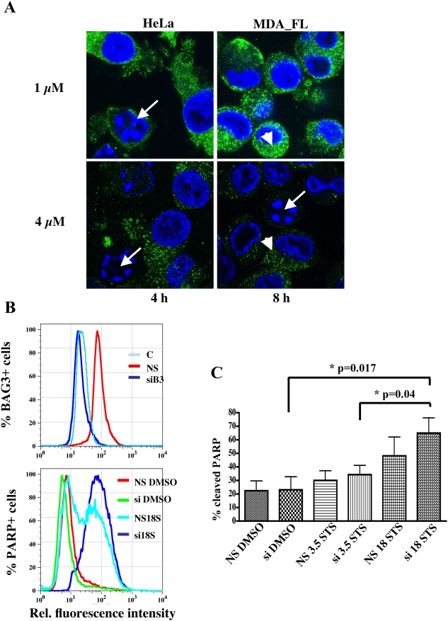
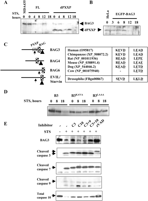
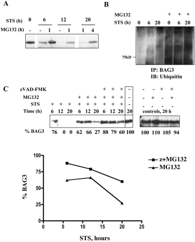
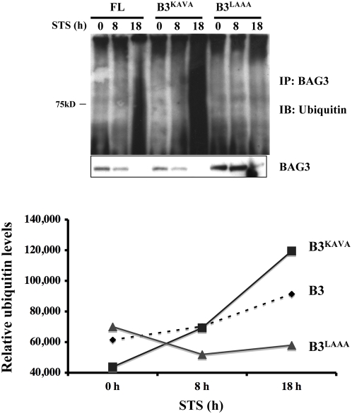
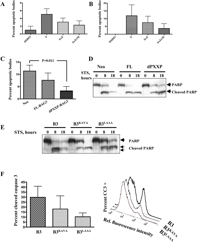
Methodology/principal findings: Staurosporine (STS) was used as a tool to test for caspase involvement in BAG3 degradation. MDA435 and HeLa human cancer cell lines exposed to STS underwent apoptosis with a concomitant time and dose-dependent loss of BAG3, suggesting the survival role of BAG3 was subject to STS regulation. zVAD-fmk or caspase 3 and 9 inhibitors provided a strong but incomplete protection of both cells and BAG3 protein. Two putative caspase cleavage sites were tested: KEVD (BAG3(E345A/D347A)) within the proline-rich center of BAG3 (PXXP) and the C-terminal LEAD site (BAG3(E516A/D518A)). PXXP deletion mutant and BAG3(E345A/D347A), or BAG3(E516A/D518A) respectively slowed or stalled STS-mediated BAG3 loss. BAG3, ubiquitinated under basal growth conditions, underwent augmented ubiquitination upon STS treatment, while there was no increase in ubiquitination of the BAG3(E516A/D518A) caspase-resistant mutant. Caspase and proteasome inhibition resulted in partial and independent protection of BAG3 whereas inhibitors of both blocked BAG3 degradation. STS-induced apoptosis was increased when BAG3 was silenced, and retention of BAG3 was associated with cytoprotection.

Conclusions/significance: BAG3 is tightly controlled by selective degradation during STS exposure. Loss of BAG3 under STS injury required sequential caspase cleavage followed by polyubiquitination and proteasomal degradation. The need for dual regulation of BAG3 in apoptosis suggests a key role for BAG3 in cancer cell resistance to apoptosis.


Conflict of interest statement

Competing Interests: The authors have declared that no competing interests exist.

Figures

Figure 1
Figure 1. STS induced apoptosis and degradation of BAG3.
A. STS induces cellular injury in a dose and time-dependent fashion. DAPI-stained STS-treated MDA435 cells show chromatin condensation consistent with apoptotic injury. Apoptotic bodies were observed with 2 and 4 µM STS at 4 hr (arrows) and there was a net loss of cells at 16 hrs. B. STS-mediated degradation of BAG3 occurs concomitant to activation of caspases 3, 7 and 9. Cleavage of caspases 3 and 9 were observed as early as 4 hrs into STS treatment. In comparison activation of caspases 8 and 10 was delayed, occurring after BAG protein loss was initiated. C, D. STS induces apoptosis in HeLa cells. Apoptotic bodies were observed in DAPI stained cells. Data points are the mean and SEM of five independent fields (n = 2). E, F. BAGs 3, 4, and 6 are lost progressively with STS treatment. Floating and adherent MDA435 cells were collected, lysed, and subjected to immunoblot. BAGs 3 and 4 (E), and 6 (F) were lost with STS exposures of 3–16 hrs. No reduction in the p50, p46, p34, or p29 BAG1 isoforms was observed (E). G. STS-mediated BAG3 degradation is neither cell line nor construct-specific. EGFP-BAG3 or EGFP-C1 empty vector were stably expressed in HeLa cells. Cells were exposed to 2 µM STS and both endogenous BAG3 and EGFP-BAG3 fusion proteins were lost over time, as early as 6 h.
Figure 2
Figure 2. Loss or silencing of BAG3 sensitizes cells to apoptosis.
A. Cells with intact nuclear morphology retain BAG3 signal. In HeLa with basal expression of BAG3, the appearance of apoptotic bodies (arrows) correlates with the loss of BAG3 green signal (arrowhead) in individual cells. In MDA_FL with forced expression of BAG3 the same phenomenon occurs although at higher STS dose and exposure time. B. PARP cleavage is augmented with BAG3 silencing and progressive STS dose. Adhered and floating MDA435 cells were stained with FITC-conjugated anti-cleaved PARP and anti BAG3 antibodies. Top graph shows BAG3 silencing by the leftward shift of the BAG3 positive population (dark blue line). Bottom graph shows the PARP+ population with BAG3 silencing and 18 h STS exposure (si 18S, dark blue line), compared with 18 h STS exposure but no silencing (NS 18STS, light blue line) along with controls (NS DMSO, si DMSO). C. Quantitation of percent apoptosis after 3.5 or 18 h of STS exposure from the cleaved PARP population (median FL-1) in response to BAG3 silencing combined with STS exposure (3.5 h and 18 h); data represent mean and SEM (n = 3).
Figure 3
Figure 3. BAG3 is a direct caspase substrate.
A, B. Deletion of the BAG3 PXXP domain delays loss of BAG3. Clonal FL-BAG3 and (d)PXXP-BAG3 MDA435 cells were treated with 2 µM STS as indicated. Cells were lysed and subjected to immunoanalysis with anti-BAG3. Delayed loss of dPXXP-BAG3, which contains putative caspase recognition sequence KEVD, is demonstrated compared with wild type protein. The delayed loss of dPXXP-BAG3 is confirmed in HeLa cells engineered to stably express EGFP-dPXXP-BAG3 by anti-EGFP (B). C. Location and alignment of putative caspase cleavage sites in BAGs 3, 4 and 6. KEVD (caspase 3 recognition site in the PXXP domain; arrow) and LEAD (caspase 9 recognition site; arrow head). Similar sites, represented to approximate scale, are found in BAGs 4 (421LELD) and 6 (998DEQD) , , but not in BAG1. These putative caspase cleavage sites are conserved in mammalian BAG proteins and in a Drosophila homolog (inset table). D. Mutation of either putative caspase cleavage site reduced loss of BAG3. Alanine mutagenesis of 344KEVD or 515LEAD was followed by stable transfection and expression. BAG3 was markedly decreased in B3 cells in response to 2 µM STS at the indicated times. BAG3KAVA cells retained much of their BAG3 protein up to 18 hr STS exposure and little or no BAG3 was lost in BAG3LAAA cells. E. Inhibition of caspases resulted in incomplete loss of BAG3. One hour pretreatment with 85 µM zVAD, followed by 2 µM STS up to 20 h, protected BAG3 as did the combination of caspase3 and 10 inhibitors; specific inhibitors concentrations were 40 µM for caspases 3 and 9, and 50 µM for caspase 10. The antibody against cleaved caspase 3 is selective to the cleaved forms and detects two active cleaved species with apparent mobility of 19 and 14 kDa. This latter species is described a further cleavage product. The antibody to caspase 10 detects only the uncleaved, total caspase 10. Arrows indicate target protein bands.
Figure 4
Figure 4. Sequential inhibition of caspases and proteasome provides collaborative protection of BAG3.
A. Proteasome inhibition provides dose-dependent partial protection of BAG3. FL-BAG3 cells were pretreated with 20 µM MG-132 for 1 or 4 hrs prior to exposure to 2 µM STS. Inhibition of proteasomal degradation by pretreatment with MG-132 prior to STS exposure reduced BAG3 degradation. B. BAG3 is ubiquitinated in response to STS treatment. FL-BAG3 cells were treated with 2 µM STS alone or following 4 hr preincubation with 20 µM MG-132. Lysates were immunoprecipitated with anti-BAG3 and the immunoblots were probed for ubiquitin. BAG3 was ubiquitinated and degraded in the absence of proteasome inhibition. C. STS-induced loss of BAG3 is minimized by the combination of caspase and proteasomal inhibition. FL-BAG3 cells were preincubated with 0.05% DMSO vehicle or 20 µM MG-132 for 4 hours with or without 1 additional hour of zVAD preincubation (85 µM). Treatments were followed by a total STS exposure of up to 20 hrs. A progressive retention of BAG3 was seen with inclusion of each inhibitor. Relative levels of BAG3, determined using ImageJ™, are indicated below the respective blots and plotted as percent remaining BAG3 with increasing STS time.
Figure 5
Figure 5. BAG3 degradation requires sequential caspase cleavage followed by ubiquitination.
Mutation of the LEAD caspase cleavage site precludes ubiquitination and loss. B3, BAG3KAVA and BAG3LAAA cells were exposed to 2 µM STS as indicated. Iodoacetamide-treated lysates were immunoprecipitated with anti-BAG3 and immunoblots probed for ubiquitin. The partial protection of BAG3KAVA results in a greater quantity of polyubiquitinated BAG3. Minimal ubiquitination of BAG3LAAA is demonstrated, consistent with a requirement for cleavage prior to ubiquitination. Quantity of polyubiquitinated BAG3 protein was estimated by densitometry.
Figure 6
Figure 6. Absence of caspase cleavage of BAG3 confers cellular protection against STS-mediated apoptosis.
A, B. STS-induced apoptosis was reduced with caspase and proteasome inhibition. Cells were pretreated with MG-132 for 4 hrs and treated with 2 µM STS and 85 µM zVAD-fmk for an additional 8 hrs. Wild type MDA435 (A) and FL-BAG3 overexpressors were fixed and stained with DAPI and apoptotic bodies scored from 3–5 independent fields; data represent mean and SEM (n = 2). C. Apoptosis is delayed in dPXXP-BAG3 lacking 344KEVD. At 8 hours of STS exposure, reduction in apoptotic bodies is seen in FL-BAG3 overexpressing cells and in dPXXP-BAG3 cells; 11–14 random fields were scored, data represent mean and SEM. D. PARP cleavage and loss of total PARP is reduced in dPXXP-BAG3 cells. STS exposure resulted in reduced PARP cleavage in FL cells compared to Neo controls, and both reduced and delayed PARP cleavage in dPXXP-BAG3 cells. E, F. Reduced apoptosis occurs in caspase-resistant BAG3 transfectants. FL-BAG3 wild type, BAG3KAVA and BAG3LAAA were treated with 2 µM STS or 0.05% DMSO vehicle control as shown. Delayed and reduced PARP cleavage is demonstrated in cells overexpressing KEVD and LEAD BAG3 mutants. Cleaved caspase-3, an earlier apoptotic event, was measured by flow cytometry after 6 h STS treatment. As with PARP cleavage, BAG3LAAA had the least cleavage of caspase 3, the right panel shows a representative histogram; data represent mean and SEM (n = 2).

Similar articles

Cited by

References

    1. Widmann C, Gibson S, Johnson GL. Caspase-dependent cleavage of signaling proteins during apoptosis. A turn-off mechanism for anti-apoptotic signals. J Biol Chem. 1998;273:7141–7147. - PubMed
    1. Karran L, Dyer MJ. Proteolytic cleavage of molecules involved in cell death or survival pathways: a role in the control of apoptosis? Crit Rev Eukaryot Gene Expr. 2001;11:269–277. - PubMed
    1. Asher G, Reuven N, Shaul Y. 20S proteasomes and protein degradation “by default”. Bioessays. 2006;28:844–849. - PubMed
    1. Takayama S, Xie Z, Reed JC. An evolutionarily conserved family of Hsp70/Hsc70 molecular chaperone regulators. J Biol Chem. 1999;274:781–786. - PubMed
    1. Lee JH, Takahashi T, Yasuhara N, Inazawa J, Kamada S, et al. Bis, a Bcl-2-binding protein that synergizes with Bcl-2 in preventing cell death. Oncogene. 1999;18:6183–6190. - PubMed

Publication types

Substances