. 2009 Aug;91(8):676-85.
doi: 10.1002/bip.21196.

Adhesion of pancreatic beta cells to biopolymer films

Affiliations

Adhesion of pancreatic beta cells to biopolymer films

S Janette Williams et al. Biopolymers. 2009 Aug.

Abstract

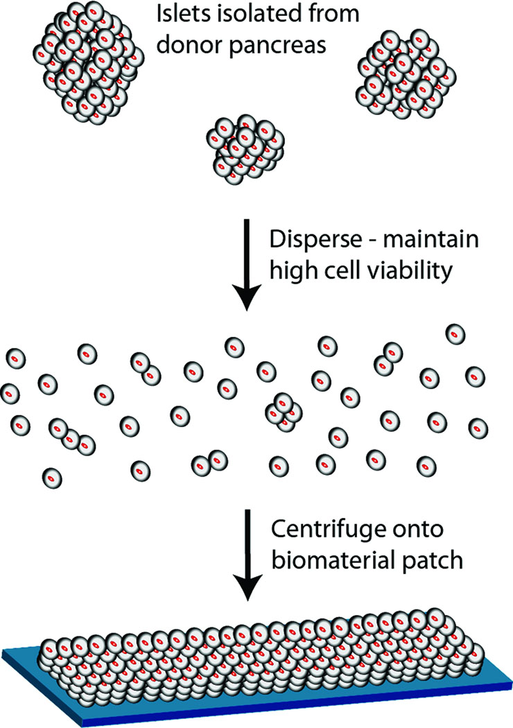
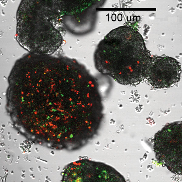
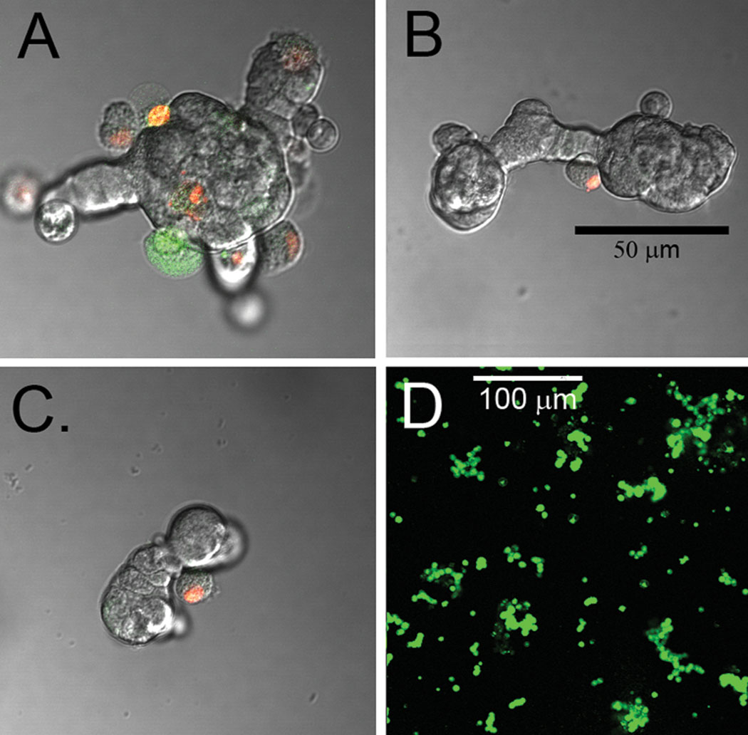
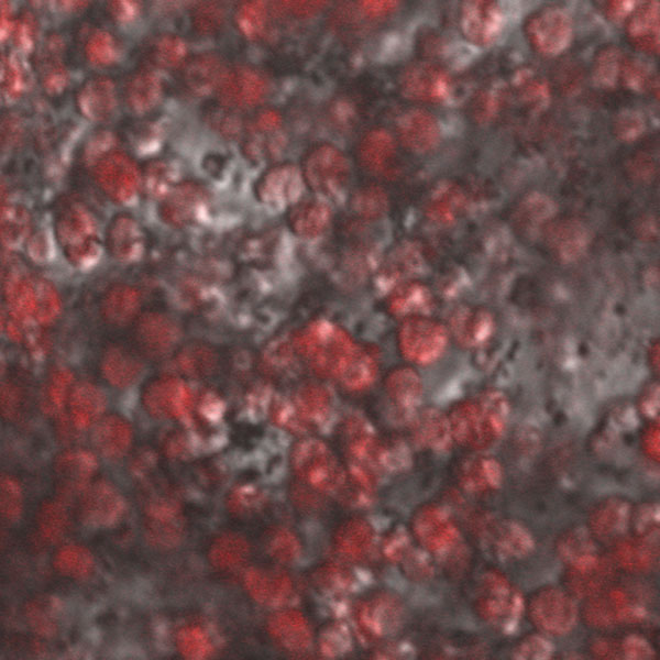
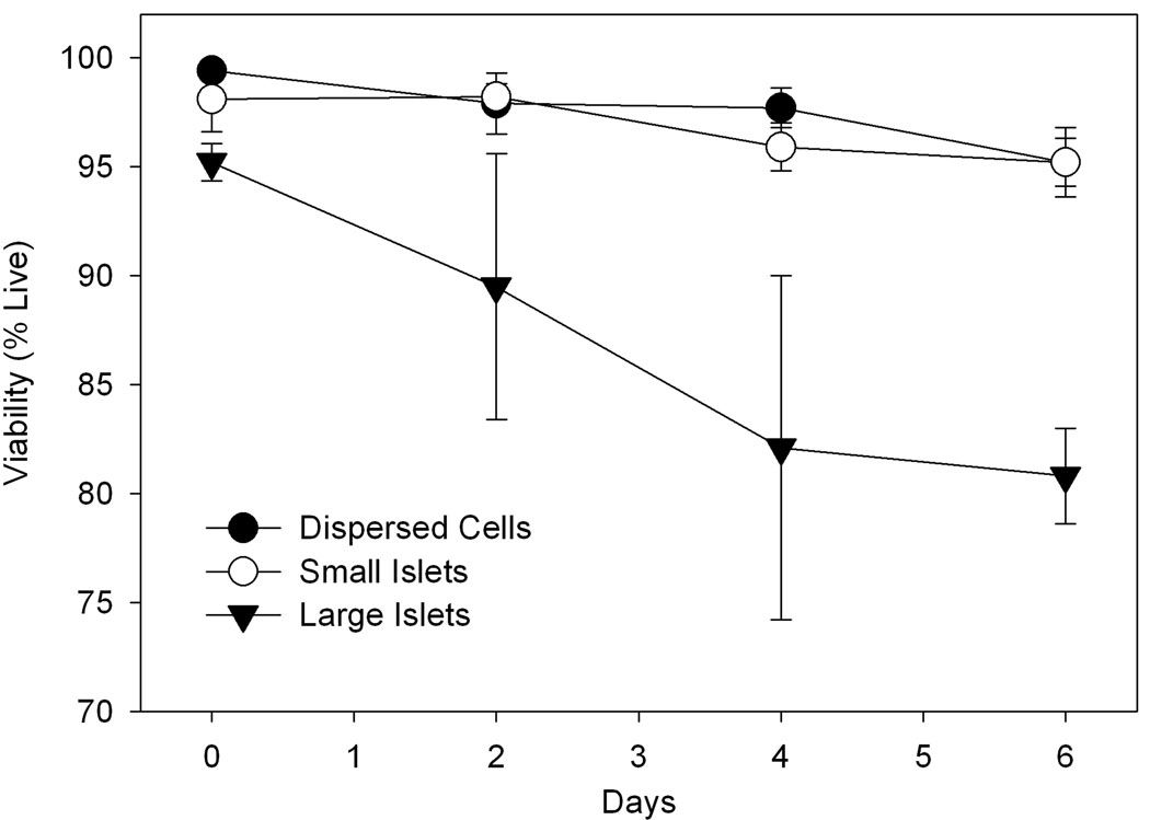
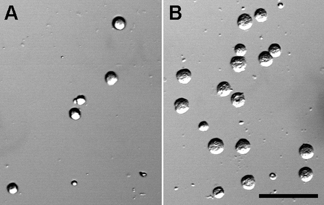
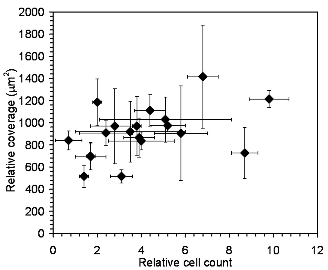
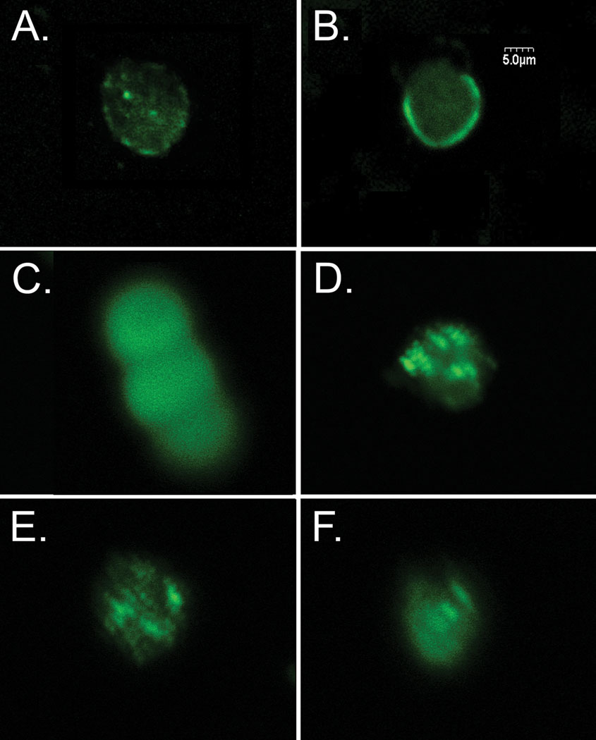
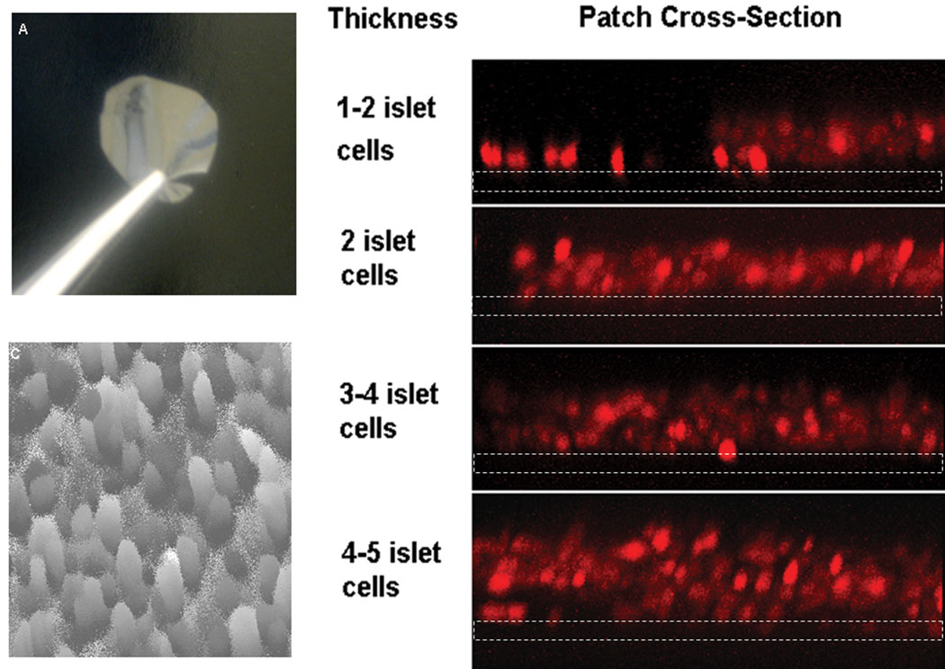
Dramatic reversal of Type 1 diabetes in patients receiving pancreatic islet transplants continues to prompt vigorous research concerning the basic mechanisms underlying patient turnaround. At the most fundamental level, transplanted islets must maintain viability and function in vitro and in vivo and should be protected from host immune rejection. Our previous reports showed enhancement of islet viability and insulin secretion per tissue mass for small islets (<125 mum) as compared with large islets (>125 mum), thus, demonstrating the effect of enhancing the mass transport of islets (i.e. increasing tissue surface area to volume ratio). Here, we report the facile dispersion of rat islets into individual cells that are layered onto the surface of a biopolymer film towards the ultimate goal of improving mass transport in islet tissue. The tightly packed structure of intact islets was disrupted by incubating in calcium-free media resulting in fragmented islets, which were further dispersed into individual or small groups of cells by using a low concentration of papain. The dispersed cells were screened for adhesion to a range of biopolymers and the nature of cell adhesion was characterized for selected groups by quantifying adherent cells, measuring the surface area coverage of the cells, and immunolabeling cells for adhesion proteins interacting with selected biopolymers. Finally, beta cells in suspension were centrifuged to form controlled numbers of cell layers on films for future work determining the mass transport limitations in the adhered tissue constructs. (c) 2009 Wiley Periodicals, Inc. Biopolymers 91: 676-685, 2009.This article was originally published online as an accepted preprint. The "Published Online" date corresponds to the preprint version. You can request a copy of the preprint by emailing the Biopolymers editorial office at biopolymers@wiley.com.


Figures

Figure 1
Figure 1
Schematic representation of the production of a multilayer beta cell construct.
Figure 2
Figure 2
Comparison of small and large rat islets isolated from the same pancreas stained for viability. The large islet on the left illustrates substantial numbers of dead cells (red and green). Small islets in the same field demonstrate a much lower percentage of dead cells.
Figure 3
Figure 3
Fragments of islets and a dispersion stained for live and dead cells. A) Fragment maden from a large islet using the enzyme dispersion method. B) Removal of calcium from the median also resulted in fragmentation of the islet. C) Utilizing both calcium depletion and enzymen resulted in smaller islet fragments. D) When islets were dispersed further via gentle pipetting,n single beta cells resulted. Figure A–C were stained with red indicating cells dead due to necrosis,n and green illustrating cell death due to apoptosis. The majority of cells dispersed into fragmentsn remained viable. Cells in D were stained with Sytox/Calcein. All green cells are viable usingn this procedure and dead cells are marked as red. The scale bar in B applies to figures A–C.
Figure 4
Figure 4
Dispersed islet cells were plated at a high density on laminin and stained with dithizone to identify beta cells.
Figure 5
Figure 5
Cell viability for cultured large (>125 µm) rat islets, small (<125 µm) rat islets, and dispersed beta cells. The decreased viability of large islets is statistically significant (p < 0.05) beyond day 3. Experiments were performed in triplicate.
Figure 6
Figure 6
A) Beta cells sparsely plated on PLGA show a higher density of attached cells after washing, indicating greater cell adhesion to the biopolymer film. B) Fewer cells remained attached to chitosan after washing.
Figure 7
Figure 7
Relative cell count values compared to the relative surface area covered by the cells (Table 2). Values are plotted with the standard deviation as error bars. Experiments were performed in triplicate.
Figure 8
Figure 8
Beta cells were plated on polymers and immunolabeled for adhesion proteins. (A) Cell plated on Pluronic® stained for E cadherin. (B) Cell on PEG immunolabeled for ZO-1. (C) Three cells on PLGA-carboxyl immunolabeled for claudin. (D) Cell on dextran sulfate immunolabeled for integrin β1 (E) Cell on chitosan immunolabeled for β catenin (F) Cell plated on dextran sulfate immunolabeled for occludin.
Figure 9
Figure 9
A) An optical image of a typical biopolymer film prior to cell deposition. B) A vertical cross-section of beta cell layers of discrete thickness, reported as the number of cell diameters 23 (beta cell diameter ~10 microns, stained using dithizone). Images were obtained by laser scanning confocal microscopy, dotted white lines depict the film location. C) An intensity plot showed that the surface topology of the top cell layer did not typically differ by more than 1 cell diameter and that complete coverage of the biopolymer substrate was attained (obtained by LSCM). Circular shapes are cells and similar shades represent similar cell height (lighter shades are higher, darker shades are lower).

References

    1. JDRF. From Research to Reality: Case Statement. 2004. Feb 06 [cited 2004 08/04/04]. Available from: www.jdrf.org.
    1. NIDDKD. Collaborative Islet Tranplant Registry, Annual Report; Sponsored by the National Institutes of Diabetes & Digestrive & Kidney Diseases; 2004. pp. 41–81.
    1. Rosenberg L, Wang R, Paraskevas S, Maysinger D. Structural and functional changes resulting from islet isolation lead to islet cell death. Surgery. 1999 Aug;126(2):393–398. - PubMed
    1. Choi SE, Choi KM, Yoon IH, Shin JY, Kim JS, Park WY, et al. IL-6 protects pancreatic islet beta cells from pro-inflammatory cytokines-induced cell death and functional impairment in vitro and in vivo. Transpl Immunol. 2004 Jun–Jul;13(1):43–53. - PubMed
    1. Wayland H. Microcirculation in pancreatic function. Microsc Res Tech. 1997 Jun 1–15;37(5–6):418–433. - PubMed

Publication types

Substances

LinkOut - more resources