. 2009 Jun 12;421(1):51-8.
doi: 10.1042/BJ20090242.

Mitochondrial peroxiredoxin 3 is more resilient to hyperoxidation than cytoplasmic peroxiredoxins

Affiliations

Mitochondrial peroxiredoxin 3 is more resilient to hyperoxidation than cytoplasmic peroxiredoxins

Andrew G Cox et al. Biochem J. .

Abstract

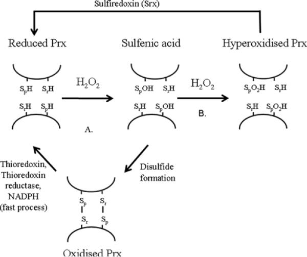
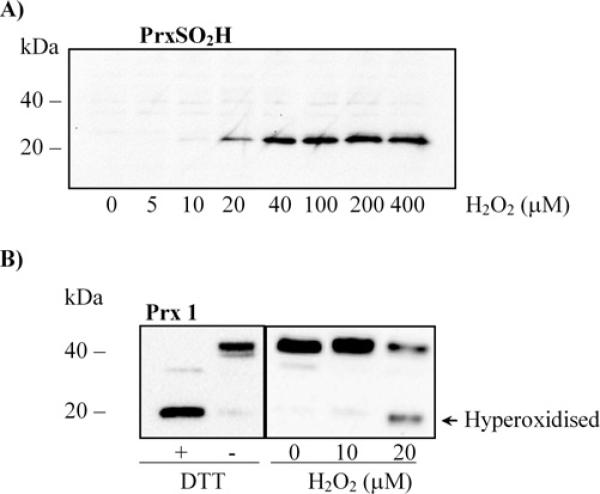
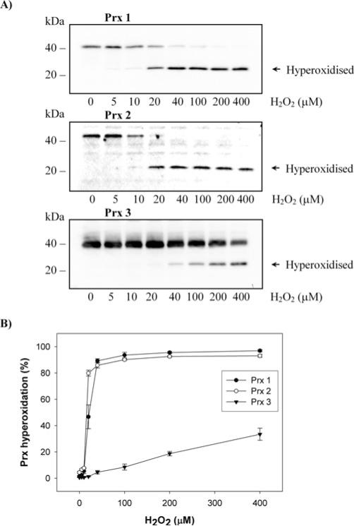
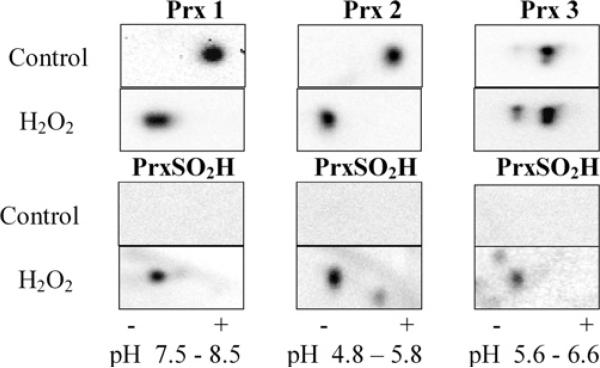
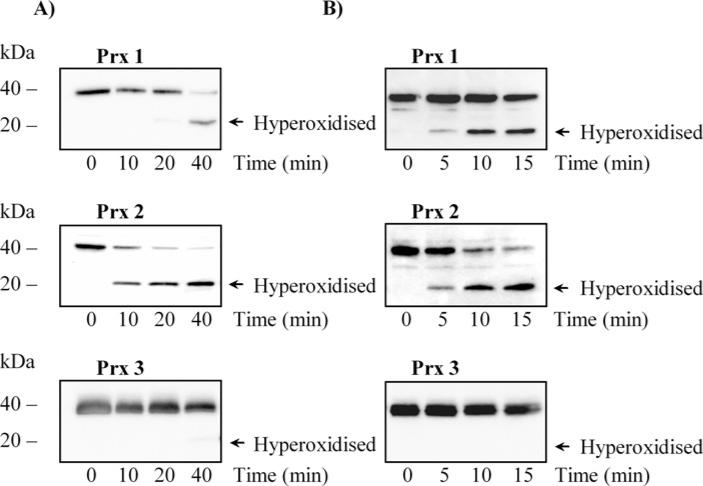
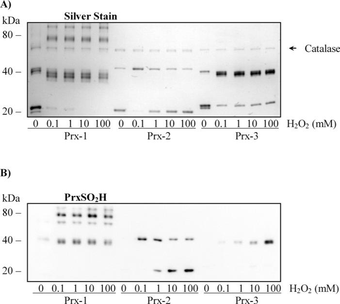
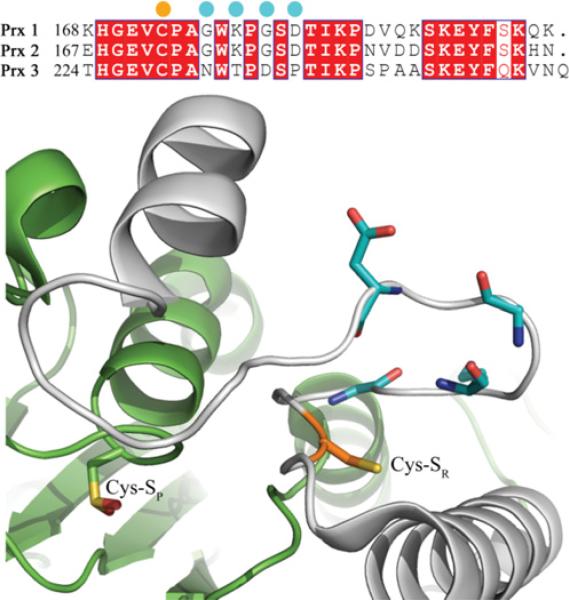
The Prxs (peroxiredoxins) are a family of cysteine-dependent peroxidases that decompose hydrogen peroxide. Prxs become hyperoxidized when a sulfenic acid formed during the catalytic cycle reacts with hydrogen peroxide. In the present study, Western blot methodology was developed to quantify hyperoxidation of individual 2-Cys Prxs in cells. It revealed that Prx 1 and 2 were hyperoxidized at lower doses of hydrogen peroxide than would be predicted from in vitro data, suggesting intracellular factors that promote hyperoxidation. In contrast, mitochondrial Prx 3 was considerably more resistant to hyperoxidation. The concentration of Prx 3 was estimated at 125 microM in the mitochondrial matrix of Jurkat T-lymphoma cells. Although the local cellular environment could influence susceptibility, purified Prx 3 was also more resistant to hyperoxidation, suggesting that despite having C-terminal motifs similar to sensitive eukaryote Prxs, other structural features must contribute to the innate resilience of Prx 3 to hyperoxidation.


Figures

Figure 1
Figure 1. Hyperoxidation during the catalytic cycle of the Prxs
(A) The peroxidatic cysteine (SpH) reacts with H2O2 to form a sulfenic acid intermediate, which can condense with the resolving cysteine on another Prx to form an intermolecular disulfide. The oxidized Prx dimer is reduced by thioredoxin to complete the catalytic cycle. (B) H2O2 can also react with the sulfenic acid intermediate to form the hyperoxidized sulfinic acid.
Figure 2
Figure 2. Expression of Prx 1, Prx 2 and Prx 3 in Jurkat cells
(A) Quantification of Prx 1, Prx 2 and Prx 3 concentration in Jurkat whole cell lysates. Jurkat cell lysates and quantities of purified Prxs were analysed by Western blotting in reducing conditions. Western blots are representative of 3 independent experiments. (B) Representative z-stack confocal images of a TMRE stained Jurkat cell (see Supplementary Movie S1 at http://www.BiochemJ.org/bj/421/bj4210051add.htm for an entire z-stack). The mitochondrial volume was calculated for each image in the z-stack. Five cells were quantified and the mitochondrial volume was calculated to be 15% (S.D. 3%).
Figure 3
Figure 3. Redox immunoblot method to monitor Prx hyperoxidation
(A) Dose-dependent increase in Prx hyperoxidation in Jurkat cells exposed to hydrogen peroxide (0–400 μM). Prx hyperoxidation was monitored by Western blotting against Prx-SO2H in non-reducing conditions. (B) Spontaneous dimerization of Prx 1 during cell harvest. Jurkat cells were harvested in extract buffer and analysed by Western blot in reducing (+DTT) or non-reducing (−DTT) conditions. Jurkat cells were exposed to hydrogen peroxide for 10 min before being harvested and examined by Western blot in non-reducing conditions. Western blots shown are representative of 3 independent experiments.
Figure 4
Figure 4. Differential susceptibility of Prxs to hyperoxidation in Jurkat cells exposed to hydrogen peroxide
(A) Jurkat cells were exposed to hydrogen peroxide (0–400 μM) for 10 min before being harvested in extract buffer and analysed by Western blot in non-reducing conditions. (B) The proportion of monomer (hyperoxidized) in each Western blot was analysed by Quantity One (Bio-Rad). Results represent the means ±S.E.M. of 3 independent experiments.
Figure 5
Figure 5. Verification of Prx hyperoxidation in Jurkat cells exposed to hydrogen peroxide
Jurkat cells were exposed to 200 μM hydrogen peroxide for 10 min before being harvested in extract buffer and resolved by 2D electrophoresis. Acidic shifts in Prx mobility were monitored by Western blot. Hyperoxidized Prx spots were validated by probing with the Prx-SO2H antibody. The isoelectric regions for each isoform were as follows, Prx 1: pH 7.5−8.5, Prx 2: pH 4.8−5.8, Prx 3: pH 5.6−6.6. Western blots shown are representative of 3 independent experiments.
Figure 6
Figure 6. Differential susceptibility of Prxs to hyperoxidation in Jurkat cells exposed to either steady state hydrogen peroxide or UV-B radiation
(A) Time course of Prx hyperoxidation in Jurkat cells exposed to a steady-state level of hydrogen peroxide (10–15 μM). (B) Timecourse of Prx hyperoxidation in Jurkat cells exposed to UV-B radiation. Cells were exposed to steady-state hydrogen peroxide or UV-B radiation for various times before being harvested with extract buffer. Samples were analysed by Western blot in non-reducing conditions. Western blots shown are representative of 3 independent experiments.
Figure 7
Figure 7. Differential susceptibility of purified Prxs to hyperoxidation
(A) Silver staining of reduced Prxs treated with hydrogen peroxide. (B) Immunodetection of Prx hyperoxidation. Reduced Prx 1, Prx 2 and Prx 3 (7 μM) were treated with hydrogen peroxide (0–100 mM) for 10 min, then 20 mM NEM and separated by SDS/PAGE under non-reducing conditions. A small amount of catalase, required to prevent re-oxidation of reduced Prxs, can be detected on the silver stain. All Prxs migrated as a monomer band under reducing conditions. A small amount of Prx-3 monomer failed to dimerize at any dose of H2O2. Protein gels and Western blots shown are representative of 3 independent experiments.
Figure 8
Figure 8. The unique C-terminal sequence of human Prx 3
The resolving Cys residue (Cys-SR) and selected Prx 3 differences are highlighted in orange and cyan, respectively. The adjacent monomer of the Prx homodimer (green) contains the peroxidatic Cys residue (Cys-SP). During normal catalysis, the C-terminus of the grey monomer rearranges to allow intermolecular disulfide bond formation. The structure shown is of human Prx 2 in the hyperoxidized state (PDB ID 1QMV) [48].

References

    1. Wood ZA, Schroder E, Harris JR, Poole LB. Structure, mechanism and regulation of peroxiredoxins. Trends Biochem. Sci. 2003;28:32–40. - PubMed
    1. Chae HZ, Chung SJ, Rhee SG. Thioredoxin-dependent peroxide reductase from yeast. J. Biol. Chem. 1994;269:27670–27678. - PubMed
    1. Rabilloud T, Heller M, Gasnier F, Luche S, Rey C, Aebersold R, Benahmed M, Louisot P, Lunardi J. Proteomics analysis of cellular response to oxidative stress – evidence for in vivo overoxidation of peroxiredoxins at their active site. J. Biol. Chem. 2002;277:19396–19401. - PubMed
    1. Chevallet M, Wagner E, Luche S, van Dorsselaer A, Leize-Wagner E, Rabilloud T. Regeneration of peroxiredoxins during recovery after oxidative stress – only some overoxidized peroxiredoxins can be reduced during recovery after oxidative stress. J. Biol. Chem. 2003;278:37146–37153. - PubMed
    1. Yang KS, Kang SW, Woo HA, Hwang SC, Chae HZ, Kim K, Rhee SG. Inactivation of human peroxiredoxin I during catalysis as the result of the oxidation of the catalytic site cysteine to cysteine-sulfinic acid. J. Biol. Chem. 2002;277:38029–38036. - PubMed

Publication types