

Noninvasive imaging of angiotensin receptors after myocardial infarction
- PMID: 19356449
- PMCID: PMC2868522
- DOI: 10.1016/j.jcmg.2007.11.007
Noninvasive imaging of angiotensin receptors after myocardial infarction
Abstract
Objectives: The purpose of this study was to evaluate the feasibility of noninvasive imaging of angiotensin II (AT) receptor upregulation in a mouse model of post-myocardial infarction (MI) heart failure (HF).
Background: Circulating AT levels do not reflect the status of upregulation of renin-angiotensin axis in the myocardium, which plays a central role in ventricular remodeling and evolution of HF after MI. Appropriately labeled AT or AT receptor blocking agents should be able to specifically target AT receptors by molecular imaging techniques.
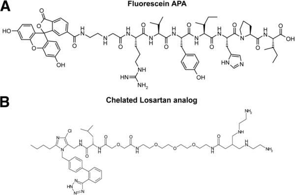
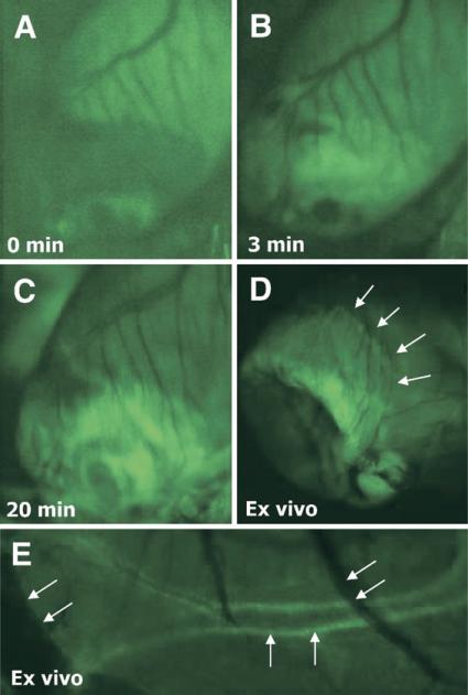
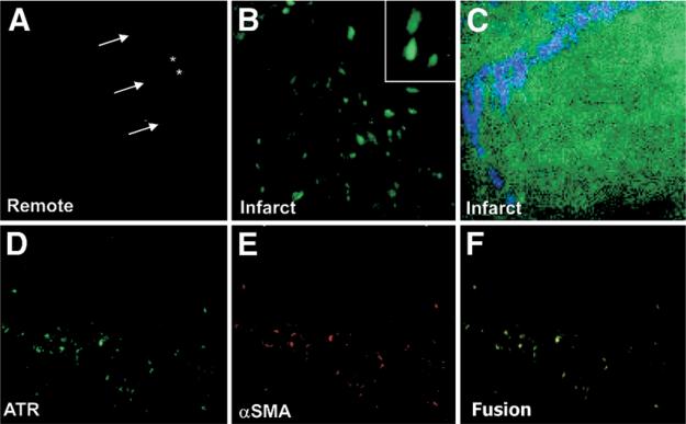
Methods: AT receptor imaging was performed in 29 mice at various time points after permanent coronary artery ligation or in controls using a fluoresceinated angiotensin peptide analog (APA) and radiolabeled losartan. The APA was used in 19 animals for intravital fluorescence microscopy on a beating mouse heart. Tc-99m losartan was used for in vivo radionuclide imaging and quantitative assessment of AT receptor expression in 10 mice. After imaging, hearts were harvested for pathological characterization using confocal and 2-photon microscopy.
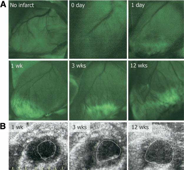
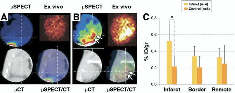
Results: No or little APA uptake was observed in control animals or within infarct regions on days 0 and 1. Distinct uptake occurred in the infarct area at 1 to 12 weeks after MI; the uptake was at maximum at 3 weeks and reduced markedly at 12 weeks after MI. Ultrasonographic examination demonstrated left ventricular remodeling, and pathologic characterization revealed localization of the APA tracer with collagen-producing myofibroblasts. Tc-99m losartan uptake in the infarct region (0.524 +/- 0.212% injected dose/g) increased 2.4-fold as compared to uptake in the control animals (0.215 +/- 0.129%; p < 0.05).
Conclusions: The present study demonstrates the feasibility of in vivo molecular imaging of AT receptors in the remodeling myocardium. Noninvasive imaging studies aimed at AT receptor expression could play a role in identification of subjects likely to develop heart failure. In addition, such a strategy could allow for optimization of anti-angiotensin therapy in patients after MI.
Figures
Comment in
-
Molecular imaging of the remodeling heart: the next step forward.JACC Cardiovasc Imaging. 2008 May;1(3):363-5. doi: 10.1016/j.jcmg.2008.03.007. JACC Cardiovasc Imaging. 2008. PMID: 19356450 No abstract available.
References
-
- Paul M, Poyan Mehr A, Kreutz R. Physiology of local renin-angiotensin systems. Physiol Rev. 2006;86:747–803. - PubMed
-
- Weber KT. Extracellular matrix remodeling in heart failure: a role for de novo angiotensin II generation. Circulation. 1997;96:4065–82. - PubMed
-
- Harada K, Sugaya T, Murakami K, Yazaki Y, Komuro I. Angiotensin II type 1A receptor knockout mice display less left ventricular remodeling and improved survival after myocardial infarction. Circulation. 1999;100:2093–9. - PubMed
-
- Pfeffer MA, Swedberg K, Granger CB, et al. Effects of candesartan on mortality and morbidity in patients with chronic heart failure: the CHARM-Overall programme. Lancet. 2003;362:759–66. - PubMed
-
- Pitt B, Poole-Wilson PA, Segal R, et al. Effect of losartan compared with captopril on mortality in patients with symptomatic heart failure: randomised trial—the Losartan Heart Failure Survival Study ELITE II. Lancet. 2000;355:1582–7. - PubMed
Publication types
MeSH terms
Substances
Grants and funding
LinkOut - more resources
Full Text Sources
Medical
Research Materials
Miscellaneous
