

Effect of chronic morphine on the dentate gyrus neurogenic microenvironment
- PMID: 19356684
- PMCID: PMC2694451
- DOI: 10.1016/j.neuroscience.2009.01.020
Effect of chronic morphine on the dentate gyrus neurogenic microenvironment
Abstract
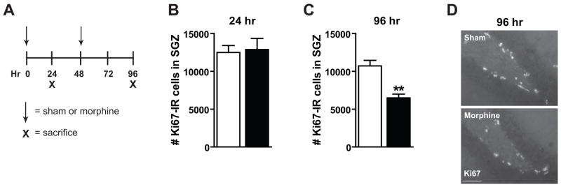
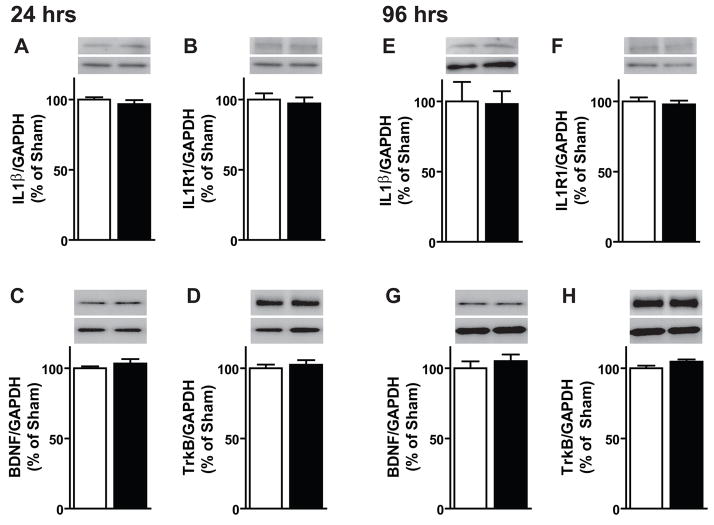
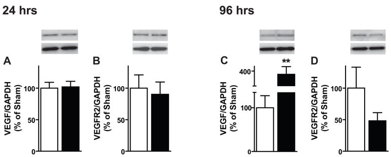
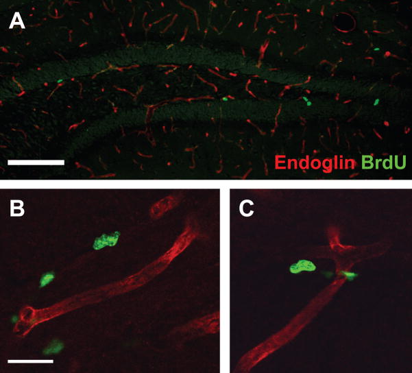
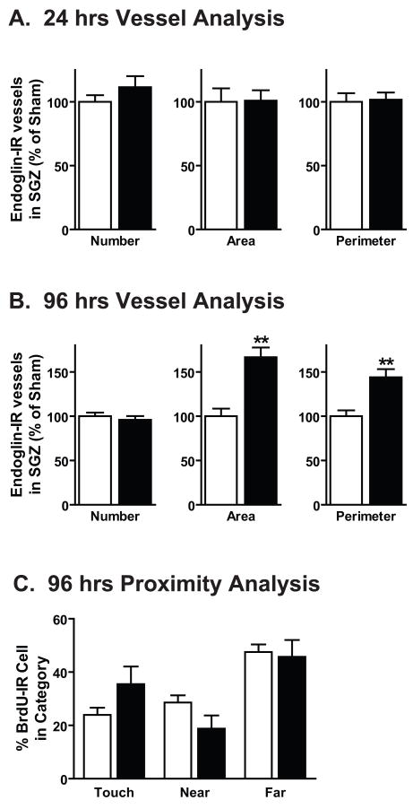
Opiates, such as morphine, decrease neurogenesis in the postnatal hippocampal subgranular zone (SGZ) by inhibiting progenitor proliferation and maturation. However, it is not known how morphine influences the growth factors and vasculature that encompass the neurogenic SGZ microenvironment. We examined morphine's effect on pro- and anti-proliferative factors in the dentate gyrus (DG; Experiment 1) as well as the DG neurovasculature itself (Experiment 2). For Experiment 1, mice were implanted with subcutaneous sham or morphine pellets (0 and 48 h) and were decapitated 24 or 96 h later. One brain hemisphere was postfixed to examine proliferation by immunohistochemistry, and a DG-enriched sample was dissected from the other hemisphere to examine the neurogenic microenvironment via immunoblotting for known pro- and anti-proliferative factors. Consistent with previous results, morphine decreased the number of proliferating cells in the SGZ, as the number of Ki67-immunoreactive (IR) cells was decreased at 96 h. Morphine did not alter DG levels of the pro-proliferative factor brain-derived neurotrophic factor, anti-proliferative factor interleukin-1 beta, or their receptors TrkB and IL1R1 at either time point. However, morphine increased the pro-proliferative factor vascular endothelial growth factor (VEGF) at 96 h. Given that VEGF is also a potent angiogenic factor, Experiment 2 examined whether the morphine-induced increase in VEGF correlated with altered DG neurovasculature. Mice were implanted with morphine pellets as in Experiment 1, and 2 h before perfusion (24 or 96 h) were administered bromodeoxyuridine (BrdU; intraperitoneal, 150 mg/kg). Tissue was co-stained for BrdU and the endothelial cell marker endoglin to enable examination of DG vessels and proximity of BrdU-IR cells to endoglin-IR vessels. At 96 h, endoglin-IR vessel area and perimeter were increased, but proximity of BrdU-IR cells to endoglin-IR vessels remained unchanged. These data suggest that following chronic morphine exposure, factors within the neurogenic microenvironment are maintained or upregulated to compensate for decreased SGZ proliferation.
Figures
References
-
- Altman J, Das GD. Autoradiographic and histological evidence of postnatal hippocampal neurogenesis in rats. J Comp Neurol. 1965;124(3):319–335. - PubMed
-
- Angelucci F, Ricci V, Pomponi M, Conte G, Mathe AA, Attilio Tonali P, Bria P. Chronic heroin and cocaine abuse is associated with decreased serum concentrations of the nerve growth factor and brain-derived neurotrophic factor. J Psychopharmacol. 2007;21(8):820–825. - PubMed
-
- Arguello AA, Harburg GC, Schonborn JR, Mandyam CD, Yamaguchi M, Eisch AJ. Time course of morphine’s effects on adult hippocampal subgranular zone reveals preferential inhibition of cells in S phase of the cell cycle and a subpopulation of immature neurons. Neuroscience. 2008;157(1):70–79. - PMC - PubMed
-
- Balasubramanian S, Ramakrishnan S, Charboneau R, Wang J, Barke RA, Roy S. Morphine sulfate inhibits hypoxia-induced vascular endothelial growth factor expression in endothelial cells and cardiac myocytes. J Mol Cell Cardiol. 2001;33(12):2179–2187. - PubMed
Publication types
MeSH terms
Substances
Grants and funding
LinkOut - more resources
Full Text Sources
