. 2009 May 29;42(8):1114-9.
doi: 10.1016/j.jbiomech.2009.02.012. Epub 2009 Apr 7.

Endothelial actin and cell stiffness is modulated by substrate stiffness in 2D and 3D

Affiliations

Endothelial actin and cell stiffness is modulated by substrate stiffness in 2D and 3D

Fitzroy J Byfield et al. J Biomech. .

Abstract

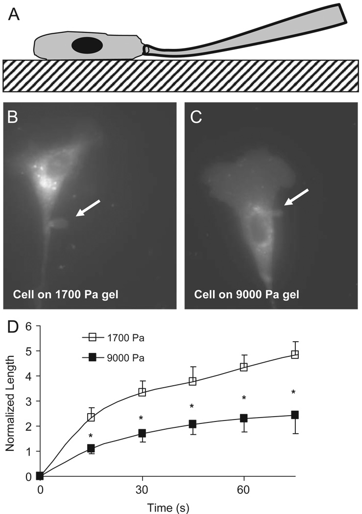
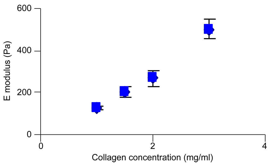
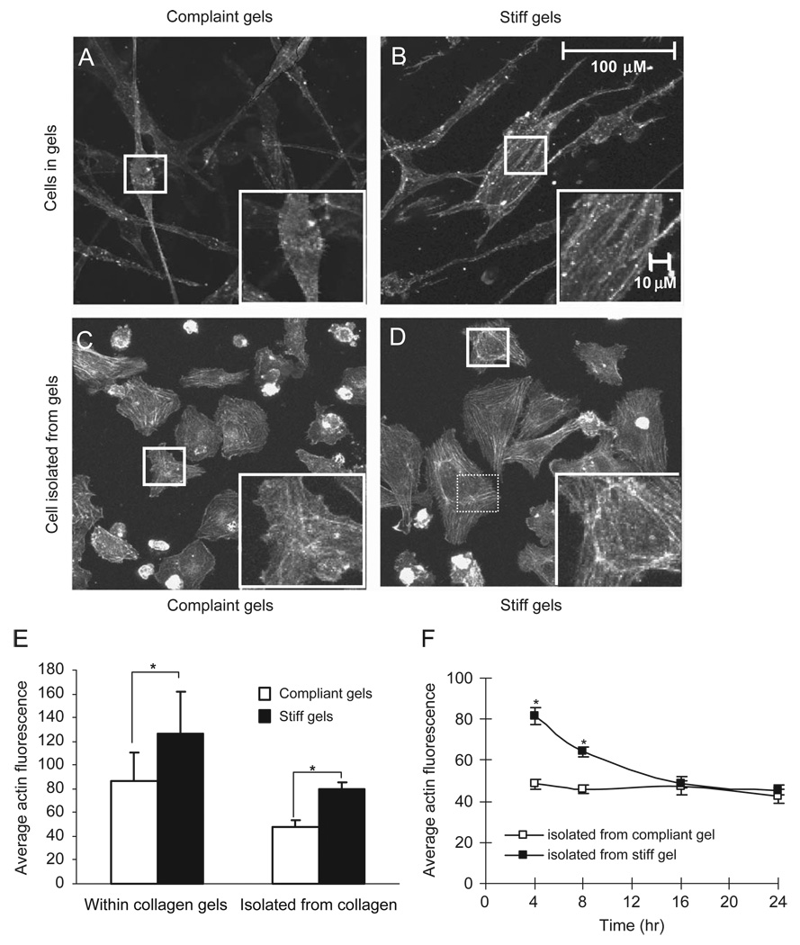
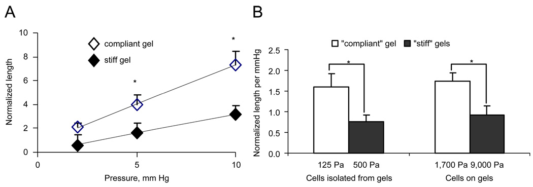
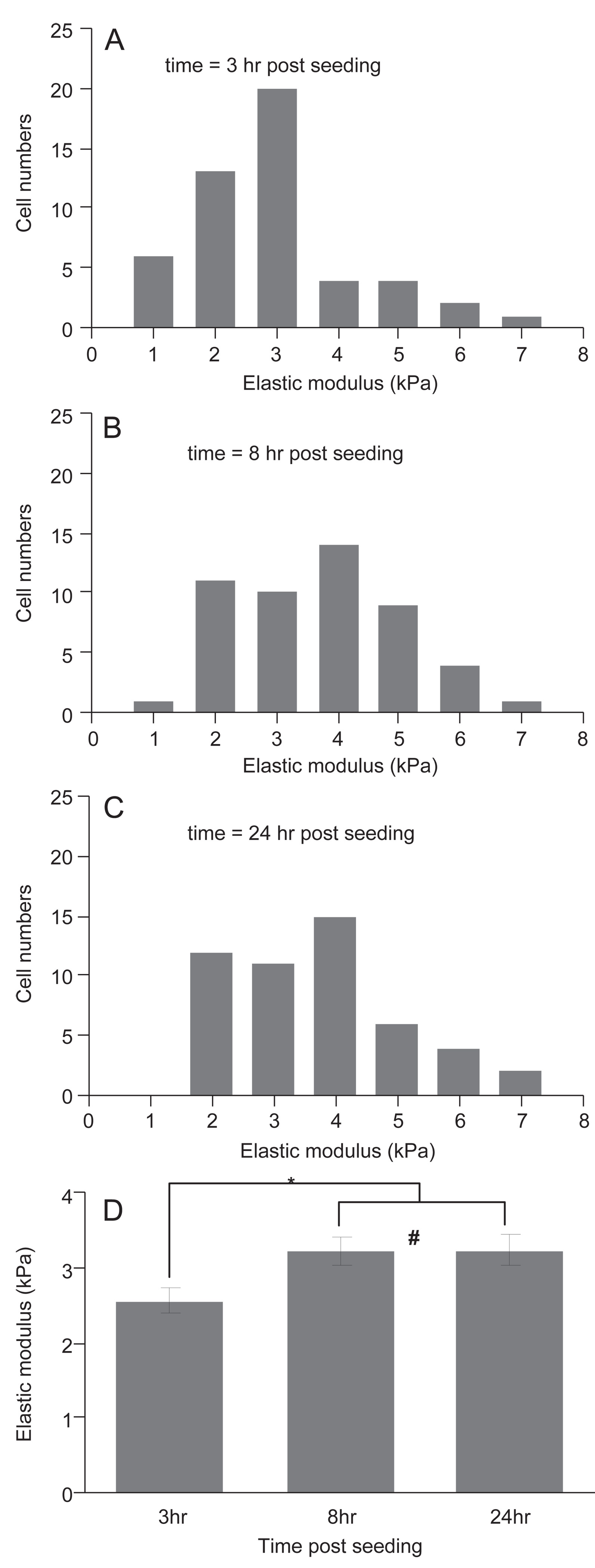
There is a growing appreciation of the profound effects that passive mechanical properties, especially the stiffness of the local environment, can have on cellular functions. Many experiments are conducted in a 2D geometry (i.e., cells grown on top of substrates of varying stiffness), which is a simplification of the 3D environment often experienced by cells in vivo. To determine how matrix dimensionality might modulate the effect of matrix stiffness on actin and cell stiffness, endothelial cells were cultured on top of and within substrates of various stiffnesses. Endothelial cells were cultured within compliant (1.0-1.5mg/ml, 124+/-8 to 202+/-27Pa) and stiff (3.0mg/ml, 502+/-48Pa) type-I collagen gels. Cells elongated and formed microvascular-like networks in both sets of gels as seen in previous studies. Cells in stiffer gels exhibited more pronounced stress fibers and approximately 1.5-fold greater staining for actin. As actin is a major determinant of a cell's mechanical properties, we hypothesized that cells in stiff gels will themselves be stiffer. To test this hypothesis, cells were isolated from the gels and their stiffness was assessed using micropipette aspiration. Cells isolated from relatively compliant gels were 1.9-fold more compliant than cells isolated from relatively stiff gels (p<0.05). Similarly, cells cultured on top of 1700Pa polyacrylamide gels were 2.0-fold more compliant that those cultured on 9000Pa (p<0.05). These data demonstrate that extracellular substrate stiffness regulates endothelial stiffness in both three- and two-dimensional environments, though the range of stiffnesses that cells respond to vary significantly in different environments.


Conflict of interest statement

Conflict of interest statement

No conflicts.

Figures

Fig. 1
Fig. 1
Microaspiration of adhered cells. The micropipette is gently pressed against the glass slide and slid into contact with the adhered cell (A). A vacuum is applied; aspirating a portion of the cell (arrow) into the pipette bore (B, C). The aspirated length is monitored as a function of time revealing that bovine aortic endothelial cells cultured on stiff substrates are more difficult to deform relative to those cultured on compliant gels (D).
Fig. 2
Fig. 2
Increasing collagen concentration increases gel stiffness. Figure shows mean values and the SEM.
Fig. 3
Fig. 3
Qualitative (A–D) and quantitative (E, F) assessment of actin for endothelial cells. Cells within compliant gels (A) exhibit less intense actin staining and less prominent stress fibers as compared to cells within stiff gels (B). These differences in actin are preserved after the cells are isolated from compliant (C) or stiff (D) gels and plated on glass slides. Quantitative image analysis (E) confirm the qualitative observation. (F) The average actin fluorescence for cells isolated from stiff and compliant gels decreases as a function of time since plating of cells onto glass slides.
Fig. 4
Fig. 4
Micropipette aspiration data for endothelial cells isolated from relatively compliant (open symbols and bars) and relatively stiff (filled symbols and bars) gels. Panel A shows the steady-state normalized aspirated length (L/a) as a function of applied vacuum. Panel B shows the normalized aspirated length per mmHg with a vacuum of 5 mmHg for both cells isolated from gels as well as those cultured on gels.
Fig. 5
Fig. 5
Elastic moduli of HUVEC following removal from a collagen gel and being plated onto glass. Histogram of elastic moduli of HUVEC measured by AFM at 3 h (A), 8 h (B), and 24 h (C) after seeding on glass slides. Average and SEM of elastic moduli as a function of time (D). * indicates p<0.02; # indicates p = 0.95.

References

    1. Byfield FJ, Aranda-Espinoza H, et al. Cholesterol depletion increases membrane stiffness of aortic endothelial cells. Biophys. J. 2004;87(5):3336–3343. - PMC - PubMed
    1. Cukierman E, Pankov R, et al. Taking cell-matrix adhesions to the third dimension. Science. 2001;294(5547):1708–1712. - PubMed
    1. Discher DE, Janmey P, et al. Tissue cells feel and respond to the stiffness of their substrate. Science. 2005;310(5751):1139–1143. - PubMed
    1. Edwards D, Gooch KJ, et al. The nucleation of receptor-mediated endocytosis. Proc. Natl. Acad. Sci. USA. 1996;93:1786–1791. - PMC - PubMed
    1. Engler A, Bacakova L, et al. Substrate compliance versus ligand density in cell on gel responses. Biophys. J. 2004;86(1 Pt 1):617–628. - PMC - PubMed

Publication types