Review
. 2009 Apr;19(2):189-96.
doi: 10.1016/j.sbi.2009.02.012. Epub 2009 Apr 6.

Class III viral membrane fusion proteins

Affiliations
Review

Class III viral membrane fusion proteins

Marija Backovic et al. Curr Opin Struct Biol. 2009 Apr.

Abstract

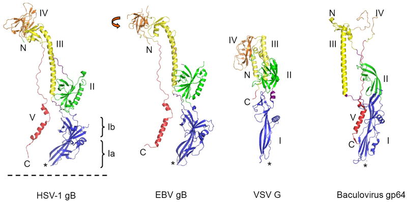
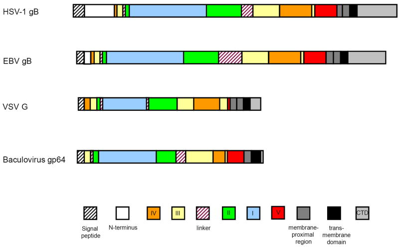
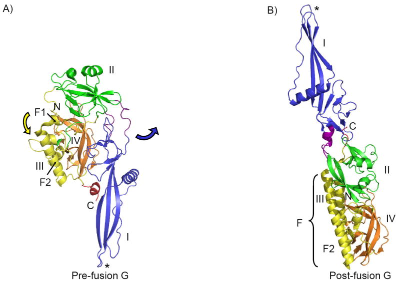
Accumulating structural studies of viral fusion glycoproteins have revealed unanticipated structural relationships between unrelated virus families and allowed the grouping of these membrane fusogens into three distinct classes. Here we review the newly identified group of class III viral fusion proteins, whose members include fusion proteins from rhabdoviruses, herpesviruses, and baculoviruses. While clearly related in structure, the class III viral fusion proteins exhibit distinct structural features in their architectures as well as in their membrane interacting fusion loops, which are likely related to their virus-specific differences in cellular entry. Further study of the similarities and differences in the class III viral fusion glycoproteins may provide greater insights into protein:membrane interactions that are key to promoting efficient bilayer fusion during virus entry.


Figures

Figure 1
Figure 1. Post-fusion conformations of class III fusion proteins
Class III fusion ectodomains form trimers, and only monomers are shown here. Domains are colored as: I (blue), II (green), III (yellow), IV (orange), V (red) and linkers (violet). N- and C-termini are marked with, respectively, letters “N” and “C”. Fusion loops are labeled with a star sign (*). C-termini and fusion loops are pointing in the same directions, as found in the post-fusion conformations of class I and class II fusion proteins. C-termini are followed by membrane-proximal regions and transmembrane domains. Dashed line represents the anticipated location of membrane. Orange arrow in EBV gB illustrates rotation of domain IV compared to the position of domain IV HSV-1 gB. Domain IV in gp64 is largely disordered.
Figure 2
Figure 2. Schematic representation of the domain organization of class III fusion proteins
Color coding of domains is the same as used in Figure 1.
Figure 3
Figure 3. Conformational change in VSV G
The ectodomain of G has been crystallized in its pre-fusion (panel A) and post-fusion (low-pH) (panel B) state. The conformational change results in flipping of domain I, carrying the fusion loops, and the C-terminus, to the opposite side of the molecule, relative to domain IV and helix F2 of domain III, which can be viewed as a rigid body and are shown in the same orientation in panels A and B. During the structural rearrangement, domains I, II and IV retain their folds, while domain III (yellow) undergoes significant refolding (central helix F2 is prolonged into the longer helix F in post-fusion form, through recruitment of helix F1, as indicated by the yellow arrow in panel A). The linker or hinge regions, which suspend domain I off the rest of the molecule (residues 47-52 and 173-180), are shown in violet. These regions undergo structural changes important for the initial stage of the conformational change, during which domain I separates from the C-terminus, swings out and rotates 94° relative to domain II (the direction of movement is indicated by the blue arrow in panel A). This is followed by repositioning of domain IV on top of domain III, and results in the more extended post-fusion conformation.

References

    1. Wilson IA, Skehel JJ, Wiley DC. Structure of the haemagglutinin membrane glycoprotein of influenza virus at 3 A resolution. Nature. 1981;289:366–373. - PubMed
    1. Rey FA, Heinz FX, Mandl C, Kunz C, Harrison SC. The envelope glycoprotein from tick-borne encephalitis virus at 2 A resolution. Nature. 1995;375:291–298. - PubMed
    1. White JM, Delos SE, Brecher M, Schornberg K. Structures and mechanisms of viral membrane fusion proteins: multiple variations on a common theme. Crit Rev Biochem Mol Biol. 2008;43:189–219. - PMC - PubMed
    1. Harrison SC. Viral membrane fusion. Nat Struct Mol Biol. 2008;15:690–698. - PMC - PubMed
    1. Kielian M, Rey FA. Virus membrane-fusion proteins: more than one way to make a hairpin. Nat Rev Microbiol. 2006;4:67–76. - PMC - PubMed

Publication types

LinkOut - more resources