. 2009 Apr;21(4):1273-90.
doi: 10.1105/tpc.107.055228. Epub 2009 Apr 7.

Interaction transcriptome analysis identifies Magnaporthe oryzae BAS1-4 as Biotrophy-associated secreted proteins in rice blast disease

Affiliations

Interaction transcriptome analysis identifies Magnaporthe oryzae BAS1-4 as Biotrophy-associated secreted proteins in rice blast disease

Gloria Mosquera et al. Plant Cell. 2009 Apr.

Abstract

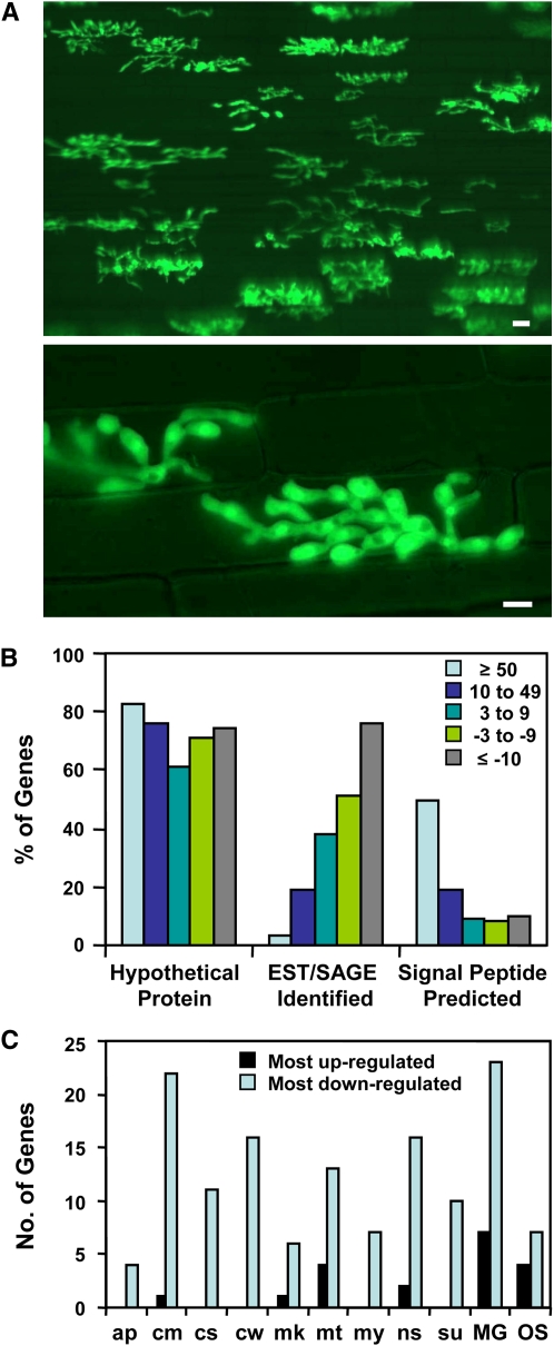
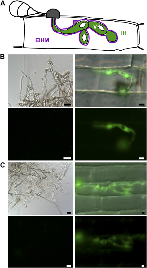
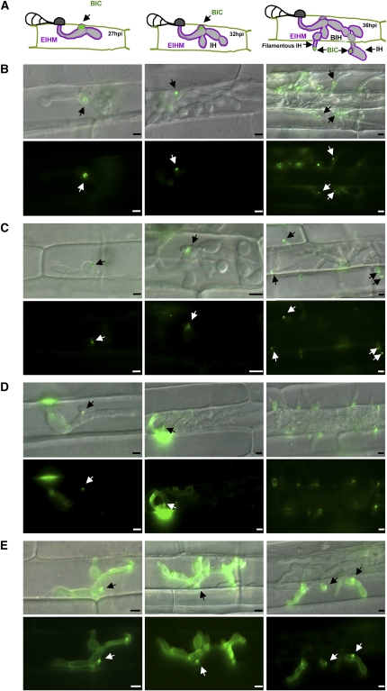
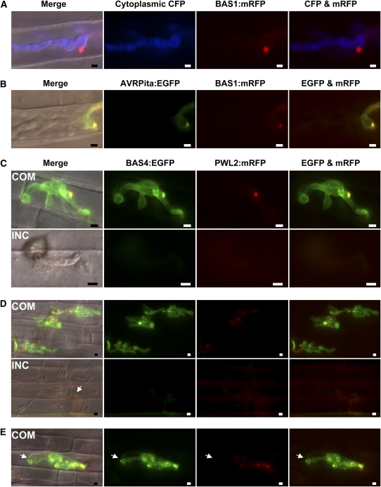
Biotrophic invasive hyphae (IH) of the blast fungus Magnaporthe oryzae secrete effectors to alter host defenses and cellular processes as they successively invade living rice (Oryza sativa) cells. However, few blast effectors have been identified. Indeed, understanding fungal and rice genes contributing to biotrophic invasion has been difficult because so few plant cells have encountered IH at the earliest infection stages. We developed a robust procedure for isolating infected-rice sheath RNAs in which approximately 20% of the RNA originated from IH in first-invaded cells. We analyzed these IH RNAs relative to control mycelial RNAs using M. oryzae oligoarrays. With a 10-fold differential expression threshold, we identified known effector PWL2 and 58 candidate effectors. Four of these candidates were confirmed to be fungal biotrophy-associated secreted (BAS) proteins. Fluorescently labeled BAS proteins were secreted into rice cells in distinct patterns in compatible, but not in incompatible, interactions. BAS1 and BAS2 proteins preferentially accumulated in biotrophic interfacial complexes along with known avirulence effectors, BAS3 showed additional localization near cell wall crossing points, and BAS4 uniformly outlined growing IH. Analysis of the same infected-tissue RNAs with rice oligoarrays identified putative effector-induced rice susceptibility genes, which are highly enriched for sensor-transduction components rather than typically identified defense response genes.


Figures

Figure 1.
Figure 1.
Biotrophic IH Are Morphologically Distinct and They Express Many Previously Undescribed Genes. (A) IH of strain KV1 growing in susceptible YT16 rice at 36 hpi. The fungus expressed cytoplasmic EYFP to facilitate microscopic assessment of infection site density before harvesting tissue for RNA preparation. Merged differential interference contrast (DIC) and fluorescence images illustrate a densely infected sheath segment (top image, bar = 20 μm) and typical IH in first-invaded epidermal cells (bottom image, bar = 5 μm). (B) Properties of pathogen genes with threefold or larger differential expression ratios, considering expression in IH relative to mycelium. Bars for each expression group indicate the percentage of the total number of genes in that group with each property. (C) Comparison of the top 50 genes expressed in IH with the bottom 50 genes for representation in EST and SAGE data sets. For each gene set, the library hits are shown for the nine genes (18%) with highest expression in IH (black bars) and the 42 genes (84%) with the lowest (light-blue bars). Many genes were identified in multiple libraries. EST libraries: ap, appressoria; cm, mycelium from complete medium; cs, conidia; cw, mycelium grown on rice cell walls; mk, pmk1 mutant; mt, mating culture; my, mycelium from minimal medium; ns, nitrogen-starved mycelium; and su, subtracted library (Ebbole et al., 2004). SAGE libraries: MG, MG_SGa, mycelium from minimal medium; OS, OSJNGg, compatible interaction at 96 hpi (Gowda et al., 2006).
Figure 2.
Figure 2.
Fungal Promoters from Upregulated Genes Are Specifically Expressed in IH. (A) Illustration showing expected IH cytoplasmic expression pattern with EGFP excluded from vacuoles (V). (B) In vitro and in planta expression of EGFP using BAS1 promoter region. (C) In vitro and in planta expression of EGFP using BAS3 promoter region. For (B) and (C), EGFP was expressed with a 1-kb promoter fragment from each gene. Left panels show phase contrast (top) and EGFP images (bottom, 3-s exposure) of mycelium, conidia, and conidiophores from agar plates (bars = 20 μm). Right panels show merged DIC and EGFP images (top) and the EGFP fluorescence alone (bottom, 3-s exposure) of IH inside rice sheath cells at 30 hpi (bars = 5 μm).
Figure 3.
Figure 3.
M. oryzae BAS Proteins Are Secreted to Distinct Locations in Sheath Epidermal Cells. (A) Illustrations showing the BIC localization pattern characteristic of AVR effectors at 27, 32, and 36 hpi. The first stage of BIC development, secretion into EIHM membranous caps at the tips of primary hyphae, is not represented. BIH, bulbous IH. (B) to (E) The promoter and coding sequence for each BAS gene (BAS1-4) was cloned with EYFP as a C-terminal translational fusion. Fungal transformants expressing BAS:EYFP fusions were observed in susceptible YT16 rice. Merged DIC and EYFP images (top) and EYFP fluorescence alone (bottom) are shown. Time points are: 27 hpi (left), 32 hpi (middle), and 36 hpi (right). BICs are indicated by arrows. Bars = 5 μm. (B) Secretion of BAS1:EYFP into BICs. (C) Secretion of BAS2:EYFP into BICs. (D) Secretion of BAS3:EYFP at 27 hpi. Note a faint BIC and fluorescence at the penetration site and outlining the primary hypha. At 32 hpi, multiple fluorescent spots were dispersed around the IH. At 36 hpi, fluorescence was near the cell wall crossing points and not in BICs at the filamentous hyphal tips. (E) Secretion of BAS4:EYFP. Fluorescence uniformly outlined the IH. Some fluorescence was also observed in BICs.
Figure 4.
Figure 4.
Fluorescent Effector PWL2 and BAS Proteins Accumulate in Susceptible YT16 but Not in Resistant Yashiro-mochi Rice. (A) BAS1:mRFP (red) is secreted into BICs and fails to colocalize with enhanced cyan fluorescent protein (CFP, blue) in the fungal cytoplasm (24 hpi in YT16). Exposure time for mRFP was 1 s and that for ECFP was 0.2 s. “Merge” corresponds to merged DIC, ECFP, and mRFP channels. Bars = 5 μm. (B) Secreted BAS1:mRFP (red) colocalizes (yellow) with AVRPita:EGFP (green) in BICs and around BIC-associated hyphal cells (YT16 at 32 hpi). Exposure times for mRFP and EGFP are 2 s. “Merge” shows DIC, EGFP, and mRFP together. Bars = 5 μm. (C) to (E) Transformants of strain O-137 (AVR-Pita1+) expressing PWL2:mRFP (red) and BAS4:EGFP (green) were inoculated on YT16 rice lacking Pi-ta (compatible [COM]) and on Yashiro-mochi containing Pi-ta (incompatible [INC]). “Merge” is DIC, EGFP, and mRFP channels together. Rice cells in (D) and (E) were plasmolyzed in 0.75 M sucrose. Exposure times: 1.5 s for mRFP and EGFP. Bars = 5 μm. (C) A COM infection site at 24 hpi has an apparently healthy IH secreting PWL2:mRFP into BICs and BAS4:EGFP around the IH. An INC site in the same experiment has a short hypha in an unhealthy rice cell. Neither fluorescent protein was observed, even with longer exposure times to detect faint signals. (D) In three COM infection sites (33 to 34 hpi), IH in plasmolyzed rice cells were secreting both PWL2:mRFP and BAS4:EGFP. In an INC site (same experiment), thin hyphae from the first-invaded cell (arrow) had moved to neighbor cells. Cytoplasm in all invaded cells was severely disrupted. Faint PWL2:mRFP fluorescence was observed, but BAS4:EGFP was not. (E) At a COM infection site (33 to 34 hpi), an IH in a plasmolyzed rice cell secreted PWL2:mRFP and BAS4:EGFP, but showed nonuniform BAS4:EGFP labeling around the IH and weak mRFP fluorescence in the BIC. BAS4:EGFP appeared to have spilled into the surrounding rice cytoplasm (arrow).

References

    1. Avilla-Adams, C., and Köller, W. (2002). Disruption of the alternative oxidase gene in Magnaporthe grisea and its impact on host infection. Mol. Plant Microbe Interact. 15 493–500. - PubMed
    1. Berruyer, R., Poussier, S., Kankanala, P., Mosquera, G., and Valent, B. (2006). Quantitative and qualitative influence of inoculation methods on in planta growth of rice blast fungus. Phytopathology 96 346–355. - PubMed
    1. Böhnert, H.U., Fudal, I., Dioh, W., Tharreau, D., Notteghem, J.-L., and Lebrun, M.-H. (2004). A putative polyketide synthase/peptide synthetase from Magnaporthe grisea signals pathogen attack to resistant rice. Plant Cell 16 2499–2513. - PMC - PubMed
    1. Bourett, T.M., Sweigard, J.A., Czymmek, K.J., Carroll, A., and Howard, R.J. (2002). Reef coral fluorescent proteins for visulaizing fungal pathogens. Fungal Genet. Biol. 37 211–220. - PubMed
    1. Campbell, R.E., Tour, O., Palmer, A.E., Steinbach, P.A., Baird, G.S., Zacharias, D.A., and Tsien, R.Y. (2002). A monomeric red fluorescent protein. Proc. Natl. Acad. Sci. USA 99 7877–7882. - PMC - PubMed

Publication types

Substances