. 2009 Jun;83(12):6135-48.
doi: 10.1128/JVI.00409-09. Epub 2009 Apr 8.

Shared and group-specific features of the rotavirus RNA polymerase reveal potential determinants of gene reassortment restriction

Affiliations

Shared and group-specific features of the rotavirus RNA polymerase reveal potential determinants of gene reassortment restriction

Sarah M McDonald et al. J Virol. 2009 Jun.

Abstract

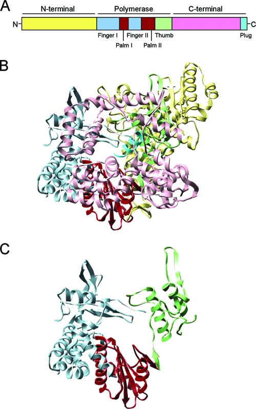
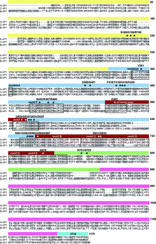
Rotaviruses (RVs) are nonenveloped, 11-segmented, double-stranded RNA viruses that are major pathogens associated with acute gastroenteritis. Group A, B, and C RVs have been isolated from humans; however, intergroup gene reassortment does not occur for reasons that remain unclear. This restriction might reflect the failure of the viral RNA-dependent RNA polymerase (RdRp; VP1) to recognize and replicate the RNA of a different group. To address this possibility, we contrasted the sequences, structures, and functions of RdRps belonging to RV groups A, B, and C (A-VP1, B-VP1, and C-VP1, respectively). We found that conserved amino acid residues are located within the hollow center of VP1 near the active site, whereas variable, group-specific residues are mostly surface exposed. By creating a three-dimensional homology model of C-VP1 with the A-VP1 crystallographic data, we provide evidence that these RV RdRps are nearly identical in their tertiary folds and that they have the same RNA template recognition mechanism that differs from that of B-VP1. Consistent with the structural data, recombinant A-VP1 and C-VP1 are capable of replicating one another's RNA templates in vitro. Nonetheless, the activity of both RdRps is strictly dependent upon the presence of cognate RV core shell protein A-VP2 or C-VP2, respectively. Together, the results of this study provide unprecedented insight into the structure and function of RV RdRps and support the notion that VP1 interactions may influence the emergence of reassortant viral strains.


Figures

FIG. 1.
FIG. 1.
Organization and structure of A-VP1. (A) Cartoon schematic illustrating the three-domain organization of A-VP1. The N-terminal domain is yellow, the C-terminal domain is pink, and the finger, palm, and thumb subdomains of the polymerase domain are pale blue, brick red, and green, respectively. The extreme C-terminal plug is cyan. (B) Ribbon representation of the A-VP1 apoenzyme crystal structure (PDB no. 2R7Q). (C) Ribbon representation of the “right-hand” A-VP1 polymerase domain. The colors in panels B and C correspond to those in panel A.
FIG. 2.
FIG. 2.
Phylogenetic relationship of A-VP1, B-VP1, and C-VP1. The dendrogram was constructed by the neighbor-joining method, systematic best tree, with gaps distributed proportionately and the Poisson correction parameter. The scale bar represents 0.2 amino acid substitution per position. The RV strain is in parentheses following the name of the organism from which it was isolated. The reovirus RdRp λ3 sequence was used as the root. Brackets indicate the VP1 group designations (A-VP1, B-VP1, and C-VP1) discussed in this report. Accession numbers are given in Materials and Methods.
FIG. 3.
FIG. 3.
Amino acid sequence alignment of A-VP1, B-VP1, and C-VP1. The primary amino acid sequence alignment of A-VP1 (strain SA11), B-VP1 (strain Cal-1), and C-VP1 (strain Bristol) is shown. Dashes indicate gaps in the protein sequence, and shading indicates conservation of amino acid identity. The domains and subdomains of A-VP1 are represented by a line above the alignment and are colored as in Fig. 1. Motifs and C-VP1 modeled residues are identified in boxed regions. Asterisks specify amino acids of interest in the present study. For a more comprehensive VP1 alignment, see Fig. S1 in the supplemental material.
FIG. 4.
FIG. 4.
Three-dimensional locations of residues conserved among A-VP1, B-VP1, and C-VP1. (A) Surface representation of the A-VP1 crystal structure (PDB no. 2R7R). Domains and subdomains are colored as in Fig. 1. Template entry and dsRNA/RNA exit tunnels are labeled for reference. The 3′CS of group A +RNA (UGUGACC) is shown (green element sticks) in the template entry tunnel, and a GTP (red element sticks) is shown in the putative cap-binding site. (B) Same image as panel A with conserved (red) and nonconserved (gray) amino acids shown. (C and D) Same image as in panels A and B but rotated 180° to the right. The NTP and template entry tunnels are shown for reference. Conserved residues (red) are shown around an unstructured loop that extends from Q346 to D357 (purple). (E and F) Same image as in panels A and B but rotated 90° to the left and shown in sagittal cutaway. All four tunnels are labeled, and free NTPs (yellow element sticks) are modeled near the active site. Conserved residues (red) are shown decorating the catalytic center and NTP and template entry tunnels.
FIG. 5.
FIG. 5.
Critical residues of the VP1 catalytic site. The image on the left is a ribbon representation of the A-VP1 polymerase domain (PDB no. 2R7R). Structural and functional motifs are colored pink (motif A), purple (motif B), green (motif C), red-orange (motif D), light yellow, (motif E), light blue (motif F), and gold (priming loop). The image on the right is a magnification of the catalytic center showing residues (colored element wires; labeled) that are conserved in A-VP1, B-VP1, and C-VP1. Nucleotides (GACC; green element sticks) of the group A +RNA are shown. Two Mg2+ ions (cyan) and free NTPs (yellow element sticks) were modeled into the priming (P) and nucleotide (N) sites based on the reovirus λ3 initiation complex (PDB no. 1MWH). For a stereo image, see Fig. S2 in the supplemental material.
FIG. 6.
FIG. 6.
Three-dimensional homology model of C-VP1. (A to D) Superimposed ribbon representations of A-VP1 (PDB no. 2R7Q; colored as in Fig. 1) and modeled C-VP1 (gray). Images are of the full-length RdRps (A), the polymerase domains with priming loops (gold; labeled) (B), the C-terminal domains (C), and the N-terminal domains (D). (E) Surface representation of A-VP1 with a ribbon diagram showing the missing flexible loop (Q346 to D327; magenta), which was modeled for C-VP1. The NTP entry tunnel is shown for reference. (F) Ribbon representation of panel E without the N- and C-terminal domains. A single A-VP1 residue (Y357; purple wire) within the modeled loop (magenta) is also conserved for the B-VP1 and C-VP1 proteins.
FIG. 7.
FIG. 7.
Surface charge distribution for A-VP1 and C-VP1. The images show the top surfaces of A-VP1 (PDB no. 2R7R) (A) and C-VP1 (B), allowing the visualization of a UGUGACC oligonucleotide (green element sticks) in the template entry tunnel. The surface is colored according to an electrostatic potential gradient, with blue and red representing positive (10 kT) and negative (−10 kT), respectively. The visible portions of the VP1 palm, N-terminal domain, and C-terminal domain are shown in a ribbon representation. The colors correspond to those in Fig. 6.
FIG. 8.
FIG. 8.
RNA recognition residues of A-VP1. Partial ribbon representation of A-VP1 (PDB no. 2R7R), revealing residues (colored element sticks, labeled) that engage the 3′CS (UGUGACC) of group A +RNA. For simplicity, the nucleotides of the RNA are numbered 1 to 7 in the 3′-to-5′ direction (i.e., C1, C2, A3, etc.). The image includes portions of the finger (pale blue), palm (purple), and thumb (green) subdomains and the N-terminal domain (yellow). Motifs B and F are labeled, and hydrogen bonds are red. Residues N186, K188, R190, F416, R701, and G702 are involved in specific recognition of the UGUG nucleotides. Residues lining the template entry tunnel (S398, S401, T418, K419, K420, G592, E593, K594, and K597) anchor the ribose-phosphate backbone of the RNA. For a stereo image, see Fig. S4 in the supplemental material.
FIG. 9.
FIG. 9.
C-VP1 has VP2-dependent in vitro activity and prefers a UGUG-containing template. (A) Purified VP1 and VP2. Recombinant A-VP1, C-VP1, A-VP2, and C-VP2 proteins were expressed in insect cells with baculovirus vectors and purified as described in Materials and Methods. Approximately 2 pmol of VP1 and 20 pmol of VP2 were electrophoresed in a 10% SDS-polyacrylamide gel and visualized by PageBlue staining. Lane M contains molecular size markers, and the values on the left are molecular sizes in kilodaltons. (B) In vitro dsRNA synthesis. Reaction mixtures included NTPs, Mg2+, Mn2+, [32P]UTP, A-VP1 or C-VP1, and a group A or C template (A-g8 +RNA or C-g9 +RNA, respectively) in the absence (none) or presence of A-VP2 or C-VP2. Radiolabeled dsRNA products were resolved in a 10% SDS-polyacrylamide gel and detected by autoradiography. (C) Template mutants. Reaction mixtures included NTPs, Mg2+, Mn2+, [32P]UTP, a group A or C protein (A-VP1/2 or C-VP1/2, respectively), and either wild-type A-g8 +RNA (UGUG) or a mutant A-g8 +RNA (AAAA) in which the VP1 recognition nucleotides UGUG were changed to AAAA. Radiolabeled dsRNA products were resolved and detected as described for panel B. The images in panels B and C are of 16-h exposures of the gels to film.

Similar articles

Cited by

References

    1. Baker, N. A., D. Sept, S. Joseph, M. J. Holst, and J. A. McCammon. 2001. Electrostatics of nanosystems: application to microtubules and the ribosome. Proc. Natl. Acad. Sci. USA 9810037-10041. - PMC - PubMed
    1. Bremont, M., P. Juste-Lesage, D. Chabanne-Vautherot, A. Charpilienne, and J. Cohen. 1992. Sequences of the four larger proteins of a porcine group C rotavirus and comparison with the equivalent group A rotavirus proteins. Virology 186684-692. - PubMed
    1. Chang, K. O., P. R. Nielsen, L. A. Ward, and L. J. Saif. 1999. Dual infection of gnotobiotic calves with bovine strains of group A and porcine-like group C rotaviruses influences pathogenesis of the group C rotavirus. J. Virol. 739284-9293. - PMC - PubMed
    1. Chen, D., C. Q. Zeng, M. J. Wentz, M. Gorziglia, M. K. Estes, and R. F. Ramig. 1994. Template-dependent, in vitro replication of rotavirus RNA. J. Virol. 687030-7039. - PMC - PubMed
    1. Chen, Z., P. R. Lambden, J. Lau, E. O. Caul, and I. N. Clarke. 2002. Human group C rotavirus: completion of the genome sequence and gene coding assignments of a non-cultivatable rotavirus. Virus Res. 83179-187. - PubMed

Publication types