. 2009 Jun;83(12):6192-8.
doi: 10.1128/JVI.00239-09. Epub 2009 Apr 8.

Acquisition of polyfunctionality by Epstein-Barr virus-specific CD8+ T cells correlates with increased resistance to galectin-1-mediated suppression

Affiliations

Acquisition of polyfunctionality by Epstein-Barr virus-specific CD8+ T cells correlates with increased resistance to galectin-1-mediated suppression

Corey Smith et al. J Virol. 2009 Jun.

Abstract

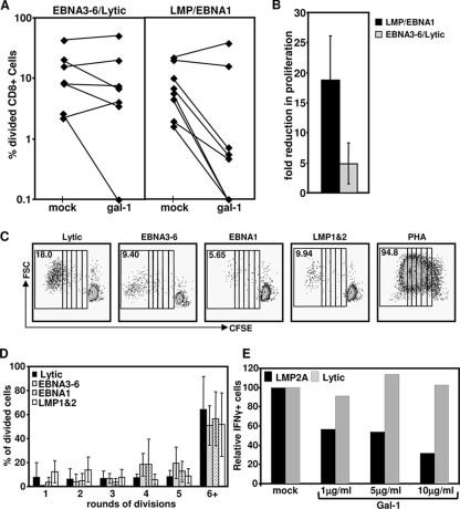
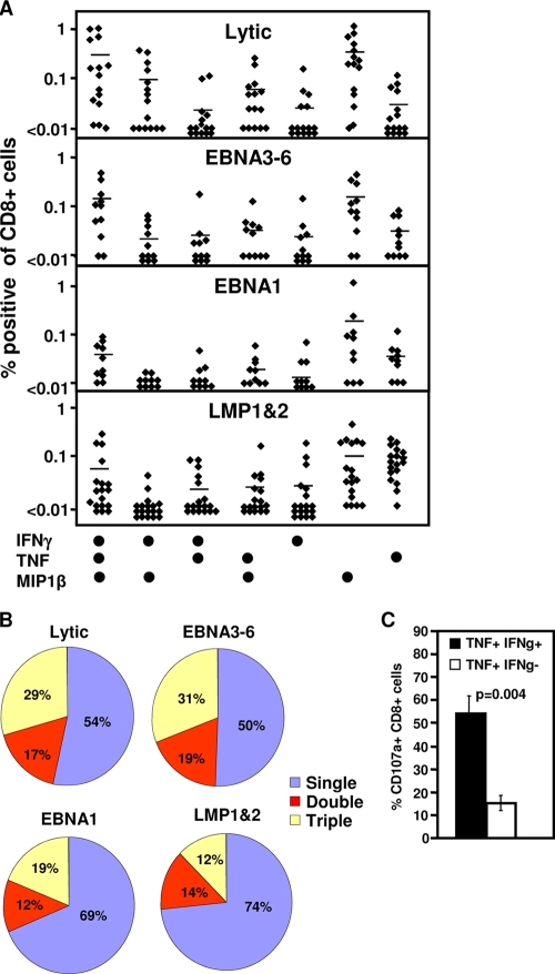
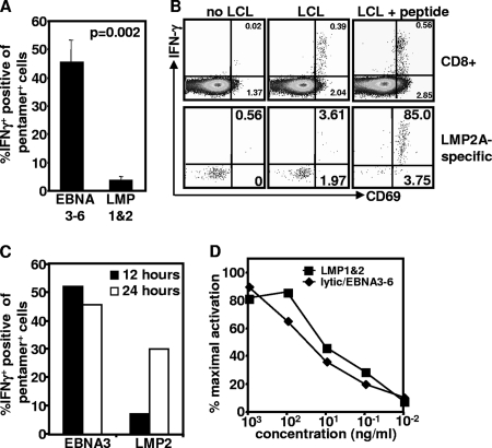
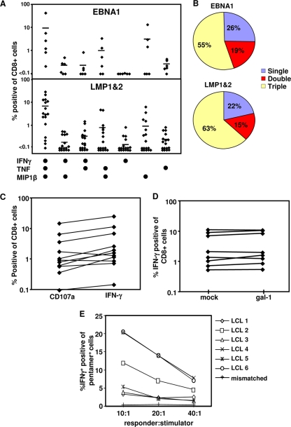
Latent membrane antigen 1 and -2 (LMP-1/2)-specific CD8(+) T cells from newly diagnosed and relapsed Hodgkin's lymphoma (HL) patients display a selective functional impairment. In contrast, CD8(+) T cells specific for Epstein-Barr virus (EBV) nuclear proteins and lytic antigens retain normal T-cell function. Reversion to a dysfunctional phenotype of LMP-1/2-specific T cells is coincident with the regression of HL. To delineate the potential basis for this differential susceptibility for the loss of function, we have carried out a comprehensive functional analysis of EBV-specific T cells using ex vivo multiparametric flow cytometry in combination with assessment of antigen-driven proliferative potential. This analysis revealed that LMP-1/2-specific T cells from healthy virus carriers display a deficient polyfunctional profile compared to that of T cells specific for epitopes derived from EBV nuclear proteins and lytic antigens. Furthermore, LMP-specific T-cells are highly susceptible to galectin-1-mediated immunosuppression and are less likely to degranulate following exposure to cognate peptide epitopes and poorly recognized endogenously processed epitopes from virus-infected B cells. More importantly, ex vivo stimulation of these T cells with an adenoviral vector encoding multiple minimal CD8(+) T-cell epitopes as a polyepitope, in combination with a gammaC cytokine, interleukin-2, restored polyfunctionality and shielded these cells from the inhibitory effects of galectin-1.


Figures

FIG. 1.
FIG. 1.
Effect of recombinant Gal-1 on the proliferation of EBV-specific T cells. PBMC from EBV-seropositive donors were labeled with CFSE, preincubated with Gal-1 at 5 μg/ml (or mock treated), and stimulated with pools of CTL epitopes of lytic cycle antigens, EBNA3-6, EBNA1, or LMP-1/2. Cells were harvested seven days later, stained with anti-CD8, and assessed for cell division on a FACSCanto. (A) Data represent the comparative proliferation in PBMC of individual donors with and without Gal-1 treatment. (B) Data represent the means and standard errors of the decreases in numbers of proliferating cells following treatment with Gal-1. (C) Representative data following stimulation with each EBV-specific peptide pool or PHA are shown. The numbers in the upper left of each panel represent the mean percentage of total proliferating CD8+ T cells following stimulation. Each box represents one round of cell division. FSC, forward scatter. (D) Data represent the means and standard deviations of the results of the rounds of division of CD8+ T cells following stimulation with the different pools of EBV-specific peptide epitopes. (E) PBMC from an EBV-seropositive donor were preincubated with Gal-1 at 1, 5, or 10 μg/ml (or mock treated) and stimulated with an LMP-2A epitope, CLGGLLTMV, or a BMLF1 (lytic antigen) epitope, GLCTLVAML, for 12 h in the presence of brefeldin A. IFN-γ expression by CD8+ T cells was assessed using intracellular cytokine assay. Data represent the percentages of IFN-γ-expressing cells relative to that in mock-treated cells.
FIG. 2.
FIG. 2.
Polyfunctionality of EBV-specific T-cell populations. (A and B) Cytokine profile of EBV-specific T-cell populations. PBMC from EBV-seropositive donors were stimulated with pools of lytic antigen, EBNA3-6, EBNA1, or LMP-1/2 epitopes for 12 h in the presence of brefeldin A. IFN-γ, TNF, and MIP1β expression by CD8+ T cells were assessed using intracellular cytokine assay. (A) Percentages of CD8+ T cells from individual donors producing each cytokine combination. (B) Average percentage of specific cells producing 1, 2, or 3 cytokines. (C) Degranulation of EBV-specific T cells. PBMC from EBV-seropositive donors were stimulated with the pools of lytic antigen, EBNA3-6, EBNA1, or LMP-1/2 epitopes for 12 h in the presence of monensin and anti-CD107α. IFN-γ (IFNg) and TNF expression by CD8+ T cells was assessed using intracellular cytokine assay. Data represent the means and standard errors of CD107α-positive cells that produce both TNF and IFN-γ or TNF alone.
FIG. 3.
FIG. 3.
Recognition of endogenous antigen by EBV-specific T cells. PBMC were stimulated with autologous LCL at a 20:1 responder-to-stimulator ratio in the presence of brefeldin A. (A) IFN-γ expression was assessed using intracellular cytokine assay. Data represent the means and standard errors of the results for IFN-γ-producing CD8+ T cells from PBMC of six donors in each group following incubation with autologous LCL for 12 h. (B) CD69 and IFN-γ expression by total CD8+ T cells or HLA pentamer-specific CD8+ T cells was assessed using intracellular cytokine assay. Representative data are from CD8+ T cells or LMP-2a-specific T cells from a single donor following stimulation with and without LCL, pulsed with and without an LMP-2a-specific epitope, CLGGLTMV. Data in the upper right quadrant are the percentages of CD69+ IFNY+ cells. Data in the lower right quadrant are the percentages of cells producing CD69 alone. (C) Representative data from single donors showing recognition of LCL by EBNA3-specific T cells recognizing the epitope FLRGRAYGL or LMP2a-specific T cells recognizing the epitope CLGGLLTMV after 12 or 24 h in the presence of brefeldin A for the previous 6 h. (D) EBV-specific T-cell avidity. PBMC from EBV-seropositive donors were stimulated overnight with 10-fold serial dilutions of HLA-matched EBV lytic antigen, EBNA3-6, EBNA1, or LMP-1/2 peptides. Activation of CD8+ T cells was assessed by the upregulation of the early activation markers CD137 and CD69.
FIG. 4.
FIG. 4.
Impact of AdE1-LMPpoly-mediated expansion on T-cell polyfunctionality. (A and B) Cytokine profiles of AdE1-LMPpoly-expanded T cells. T cells from cultures stimulated with AdE1-LMPpoly were incubated with EBNA1 or LMP-1/2 epitopes in the presence of brefeldin A. IFN-γ, TNF, and MIP1β expression by CD8+ T cells was assessed using intracellular cytokine assay. (A) Percentages of CD8+ T cells from individual donors producing each cytokine combination. (B) Mean percentages of specific cells producing 1, 2, or 3 cytokines. (C) Degranulation of AdE1-LMPpoly-stimulated T cells. Cultured T cells were incubated with EBNA1 or LMP-1/2 epitopes in the presence of monensin and anti-CD107α. Data represent the frequency of T cells that produce CD107α or IFN-γ in response to LMP or EBNA1 CTL epitopes. (D) Effect of recombinant Gal-1 on the proliferation of AdE1-LMPpoly-expanded T cells. AdE1-LMPpoly-expanded T cells were preincubated with Gal-1 at 5 μg/ml (or mock treated) and stimulated with LMP-1/2 CTL epitopes. IFN-γ expression by CD8+ T cells was assessed using intracellular cytokine assay. Data represent the comparative proliferation in PBMC from individual donors with and without Gal-1 treatment. (E) AdE1-LMPpoly-stimulated T cells were stimulated with autologous LCL at responder-to-stimulator ratios of 10:1, 20:1, and 40:1 in the presence of brefeldin A for 12 h. Data represent the percentages of IFN-γ-producing, MHC peptide pentamer-specific T cells following incubation with a panel of HLA-matched LCLs.

Similar articles

Cited by

References

    1. Almeida, J. R., D. A. Price, L. Papagno, Z. A. Arkoub, D. Sauce, E. Bornstein, T. E. Asher, A. Samri, A. Schnuriger, I. Theodorou, D. Costagliola, C. Rouzioux, H. Agut, A. G. Marcelin, D. Douek, B. Autran, and V. Appay. 2007. Superior control of HIV-1 replication by CD8+ T cells is reflected by their avidity, polyfunctionality, and clonal turnover. J. Exp. Med. 2042473-2485. - PMC - PubMed
    1. Badr, G., N. Bedard, M. S. Abdel-Hakeem, L. Trautmann, B. Willems, J. P. Villeneuve, E. K. Haddad, R. P. Sekaly, J. Bruneau, and N. H. Shoukry. 2008. Early interferon therapy for hepatitis C virus infection rescues polyfunctional, long-lived CD8+ memory T cells. J. Virol. 8210017-10031. - PMC - PubMed
    1. Gandhi, M. K., E. Lambley, J. Duraiswamy, U. Dua, C. Smith, S. Elliott, D. Gill, P. Marlton, J. Seymour, and R. Khanna. 2006. Expression of LAG-3 by tumor-infiltrating lymphocytes is coincident with the suppression of latent membrane antigen-specific CD8+ T-cell function in Hodgkin lymphoma patients. Blood 1082280-2289. - PubMed
    1. Gandhi, M. K., G. Moll, C. Smith, U. Dua, E. Lambley, O. Ramuz, D. Gill, P. Marlton, J. F. Seymour, and R. Khanna. 2007. Galectin-1 mediated suppression of Epstein-Barr virus specific T-cell immunity in classic Hodgkin lymphoma. Blood 1101326-1329. - PMC - PubMed
    1. Guppy, A. E., E. Rawlings, J. A. Madrigal, P. L. Amlot, and L. D. Barber. 2007. A quantitative assay for Epstein-Barr virus-specific immunity shows interferon-gamma producing CD8+ T cells increase during immunosuppression reduction to treat posttransplant lymphoproliferative disease. Transplantation 841534-1539. - PubMed

Publication types

MeSH terms