. 2009 Jun;296(6):F1279-90.
doi: 10.1152/ajprenal.00013.2009. Epub 2009 Apr 8.

RhCG is the major putative ammonia transporter expressed in the human kidney, and RhBG is not expressed at detectable levels

Affiliations

RhCG is the major putative ammonia transporter expressed in the human kidney, and RhBG is not expressed at detectable levels

Alice C N Brown et al. Am J Physiol Renal Physiol. 2009 Jun.

Abstract

Rhesus glycoprotein homologs RhAG, RhBG, and RhCG comprise a recently identified branch of the Mep/Amt ammonia transporter family. Animal studies have shown that RhBG and RhCG are present in the kidney distal tubules. Studies in mouse and rat tissue suggest a basolateral localization for RhBG in cells of the distal tubules including the alpha-intercalated cells (alpha-IC), but no localization of RhBG has been reported in human tissue. To date RhCG localization has been described as exclusively apical plasma membrane in mouse and rat kidney, or apical and basolateral in humans, and some mouse and rat tissue studies. We raised novel antibodies to RhBG and RhCG to examine their localization in the human kidney. Madin-Darby canine kidney (MDCKI) cell lines stably expressing human green fluorescent protein-tagged RhBG or RhCG and human tissue lysates were used to demonstrate the specificity of these antibodies for detecting RhBG and RhCG. Using immunoperoxidase staining and antigen liberation techniques, both apical and basolateral RhCG localization was observed in the majority of the cells of the distal convoluted tubule and IC of the connecting tubule and collecting duct. Confocal microscopic imaging of normal human kidney cryosections showed that RhCG staining was predominantly localized to the apical membrane in these cells with some basolateral and intracellular staining evident. A proportion of RhCG staining labeled kAE1-positive cells, confirming that RhCG is localized to the alpha-IC cells. Surprisingly, no RhBG protein was detectable in the human kidney by Western blot analysis of tissue lysates, or by immunohistochemistry or confocal microscopy of tissue sections. The same antibodies, however, could detect RhBG in rat tissue. We conclude that under normal conditions, RhCG is the major putative ammonia transporter expressed in the human kidney and RhBG is not expressed at detectable levels.


Figures

Fig. 1.
Fig. 1.
Polarized localization of green fluorescent protein (GFP)-tagged Rh glycoprotein homolog (RhBG) and GFP-RhCG in Madin-Darby canine kidney (MDCKI) stable cell lines. MDCKI cells stably transfected with NH2 terminally GFP-tagged RhCG (A) or RhBG (B) were polarized on filters. Cells were stained with rabbit anti-GFP and mouse anti-Na+-K+-ATPase antibodies and then visualized with anti-rabbit Alexa 488 (green) and anti-mouse Alexa 594 antibodies. Images were taken in a subapical plane both parallel (xy) and perpendicular to the cells (along the white line in the xy panel, xz). Merged images of the perpendicular sections demonstrate the majority of RhCG is apically localized, and RhBG has a nonpolarized localization. Bar = 20 μm.
Fig. 2.
Fig. 2.
Novel RhBG and RhCG antibodies are specific to GFP-RhBG or GFP-RhCG when analyzed by Western blotting. MDCKI cells stably expressing GFP-tagged RhBG or RhCG were treated with sodium butyrate to increase protein expression, lysed, and immunoblotted with polyclonal antibodies to GFP (A), αRhCG-CT1 (B), αRhCG-CT2 (C), αRhBG-CT1 (D), αRhBG-NT (E), and αRhBG-CT (F). All RhBG and RhCG antibodies bind specifically to the Rh glycoprotein against which they were raised.
Fig. 3.
Fig. 3.
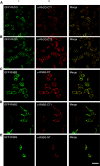
Novel RhBG and RhCG antibodies are specific for GFP-RhBG or GFP-RhCG in immunofluorescence experiments. MDCKI cells stably expressing NH2 terminally GFP-tagged RhCG (A and B) or RhBG (CE) were fixed and immunolabeled with polyclonal antibodies αRhCG-CT1 (A), αRhCG-CT2 (B), αRhBG-CT (C), αRhBG-CT1 (D), or αRhBG-NT (E). GFP-RhBG or GFP-RhCG expression was visualized by GFP fluorescence (column 1) and RhBG/RhCG antibody binding by anti-rabbit Alexa 594 secondary antibody (column 2). Bar = 50 μm.
Fig. 4.
Fig. 4.
Novel antibodies to RhCG bound specifically to human RhCG when human and rat kidney lysate was analyzed by Western blotting. Fifty micrograms of human or rat renal cortex lysate were loaded, subjected to SDS PAGE, and analyzed by immunoblotting. Anti-RhCG antibodies, αRhCG-CT1 (A) and αRhCG-CT2 (B), detect a ∼52- to 55-kDa polypeptide, which for αRhCG-CT1 was specifically competed by addition of 1 mg/ml of immunizing peptide to αRhCG-CT1 before incubation with the membrane (A). Blots were also incubated with antibodies against human erythrocyte membrane protein anion exchanger 1 (kAE1; Bric170; C) and β-actin (D) to demonstrate loading of human and rat samples.
Fig. 5.
Fig. 5.
Immunoperoxidase staining of a human kidney with rabbit αRhCG-CT1 antibody and various markers of parts of the tubule. A: control section of cortex stained by αRhCG-CT1 preincubated with the immunizing peptide. B: low-magnification view of the cortex (c) and outer medulla (m). Glomeruli and proximal tubules are not stained. Only some parts of tubules are detected, and the staining appears mostly continuous in the cortex and intermittent in the medulla. The arrows indicate parts of tubules that show staining for RhCG-CT1 but not for uromodulin in an adjacent section (C) or vice versa. RhCG is therefore not expressed in the thick limb of the loop of Henle. D: high-magnification image of the cortex to show that in some tubules every cell expresses RhCG, and the arrows indicate that the apical membrane is most strongly stained, although there is also staining of the basolateral membrane and to a lesser extent the cytoplasm. E: high-magnification image of the inner medulla to show that only some cells in collecting ducts (cd) are stained, mainly on the basolateral membrane (arrowed). In F there is a group of tubules expressing RhCG, and G shows that the same tubules in an adjacent section express the sodium-chloride cotransporter on their apical membrane (arrows). This confirms expression of RhCG in the distal convoluted tubule. In H, RhCG-CT1 immunostaining is seen in cortical tubules not only continuously as in C and F, but also discontinuously (arrows). Adjacent sections show that the discontinuous part expresses the amiloride-sensitive epithelial sodium channel (I) and that both parts express 11β-hydroxysteroid dehydrogenase type 2 (J). This shows that the continuous staining of the distal convoluted tubule changes to staining only of intercalated cells in the connecting tubule. In K, a connecting tubule has RhCG-CT1 staining only in intercalated cells, while in an adjacent section principal cells express aquaporin-2 on their apical membrane (arrows; L).
Fig. 6.
Fig. 6.
RhBG is detectable on Western blot of rat kidney lysate but not of healthy human kidney lysate. Fifty micrograms of each lysate were prepared as in Fig. 4. Two COOH-terminal antibodies, αRhBG-CT (A) and affinity-purified αRhBG-CT1 (B) bind a ∼54-kDa polypeptide in rat cortex tissue that can be specifically competed by preincubation of the antibodies with 1 mg/ml of immunizing peptide. Neither antibody detects an equivalent polypeptide in human tissue that can be specifically competed. The NH2-terminal RhBG antibody, αRhBG-NT, binds nonspecifically in both rat and human tissue with equivalent background bands detected in the presence or absence of immunizing peptide (C).
Fig. 7.
Fig. 7.
RhBG is not detectable by immunoperoxidase stain on human renal cortex, renal medulla, or liver sections but is detectable on immunoperoxidase-stained rat kidney. Human renal cortex and medulla sections were stained with αRhBG-CT (A and B) and αRhBG-CT1 (C and D) at a high concentration (1:50). There is no reactivity to RhBG using αRhBG-CT in any region of the cortex (A) or medulla (B) and only a faint cytoplasmic staining in thick ascending limbs using αRhBG-CT1 (C and D). Preincubation of αRhBG-CT1 with immunizing peptide did remove this antibody reactivity (E). Rat renal cortex immunostained with αRhBG-CT showed basolateral RhBG staining in the distal tubules (F). Human liver sections were first stained with αRhCG-CT1 antibody (G). A few scattered hepatocytes show strong membrane staining (G), which is removed following preincubation of αRhCG-CT1 antibody with immunizing peptide (H). Liver immunostained with αRhBG-CT1 showed no reactivity in any cell type (I).
Fig. 8.
Fig. 8.
RhCG but not RhBG can be detected by immunofluorescence staining of healthy human renal cortex sections. Renal cortex sections of a human kidney were costained with rabbit αRhCG-CT1 antibody (column 1, A and B) and a mouse antibody to α-intercalated cell (IC) basolaterally localized protein kAE1 (column 2). Immunofluorescence visualization using anti-rabbit Alexa 488 and anti-mouse Alexa 594 antibodies show RhCG and kAE1 costain α-IC, where RhCG is predominantly apically localized and kAE1 is basolaterally localized (A). Preincubation with immunizing peptide competed RhCG binding; hence staining of the tissue for RhCG was no longer observed (B). Human renal cortex sections were also stained for RhBG, using αRhBG-CT (A), affinity-purified αRhBG-CT1 (B) and αRhBG-NT (C) antibodies, and costained for kAE1 using Bric170. No RhBG immunoreactivity was observed with any of the three RhBG antibodies in any region or cell type of the kidney cortex including the kAE1-positive α-IC. This result was observed in several independent tissue samples. Bar = 50 μm.

References

    1. Bakouh N, Benjelloun F, Hulin P, Brouillard F, Edelman A, Cherif-Zahar B, Planelles G. NH3 is involved in the NH4+ transport induced by the functional expression of the human Rh C glycoprotein. J Biol Chem 279: 15975–15983, 2004. - PubMed
    1. Biner HL, Arpin-Bott MP, Loffing J, Wang X, Knepper M, Hebert SC, Kaissling B. Human cortical distal nephron: distribution of electrolyte and water transport pathways. J Am Soc Nephrol 13: 836–847, 2002. - PubMed
    1. Biver S, Belge H, Bourgeois S, Van Vooren P, Nowik M, Scohy S, Houillier P, Szpirer J, Szpirer C, Wagner CA, Devuyst O, Marini AM. A role for Rhesus factor Rhcg in renal ammonium excretion and male fertility. Nature 456: 339–343, 2008. - PubMed
    1. Bostanjoglo M, Reeves WB, Reilly RF, Velazquez H, Robertson N, Litwack G, Morsing P, Dorup J, Bachmann S, Ellison DH. 11Beta-hydroxysteroid dehydrogenase, mineralocorticoid receptor, and thiazide-sensitive Na-Cl cotransporter expression by distal tubules. J Am Soc Nephrol 9: 1347–1358, 1998. - PubMed
    1. Bruce LJ, Beckmann R, Ribeiro ML, Peters LL, Chasis JA, Delaunay J, Mohandas N, Anstee DJ, Tanner MJ. A band 3-based macrocomplex of integral and peripheral proteins in the RBC membrane. Blood 101: 4180–4188, 2003. - PubMed

Publication types

LinkOut - more resources