

Comparative Study
doi: 10.1523/JNEUROSCI.0035-09.2009.
An oligovascular niche: cerebral endothelial cells promote the survival and proliferation of oligodendrocyte precursor cells
Affiliations
- PMID: 19357263
- PMCID: PMC3849498
- DOI: 10.1523/JNEUROSCI.0035-09.2009
Item in Clipboard
Comparative Study
An oligovascular niche: cerebral endothelial cells promote the survival and proliferation of oligodendrocyte precursor cells
J Neurosci.
.
Abstract
We show that cerebral endothelial cells secrete trophic factors that support the survival and proliferation of rat oligodendrocyte precursor cells (OPCs). This OPC-supportive phenomenon was mediated by Akt and Src signaling pathways. Noncytotoxic levels of oxidative stress downregulate trophic factor production and disrupt the ability of cerebral endothelial cells to support OPCs. These data suggest that a novel oligovascular niche may be important for sustaining oligodendrocyte renewal and homeostasis in mammalian brain.
Figures
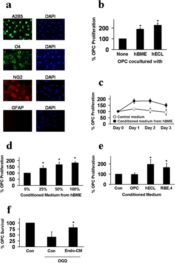
a, Immunostaining demonstrated that OPCs expressed A2B5 (green), O4 (green), NG2 (red), and lacked GFAP (red). Nuclei were stained with DAPI (blue). b, Coculturing with cerebral endothelial cells promoted OPC proliferation. Two different types of endothelial cells were tested, primary human brain microvascular endothelial cells (hBME) and a human brain endothelial cell line (hECL). c, Conditioned media from hBME amplified OPC proliferation over 1–3 d in culture. d, Different dilutions of hBME-conditioned media showed a “dose–response” effect on OPC proliferation. e, Conditioned media from a rat brain endothelial cell line (RBE.4) equally promoted OPC proliferation compared with media from a human brain endothelial cell line (hECL). No effects were seen with conditioned media from matching OPC cultures. f, Conditioned media from hBME also showed OPC-protective effects against oxygen–glucose deprivation (OGD)-induced stress. *p < 0.05.
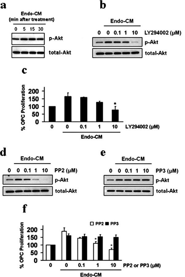
a, Conditioned media from human brain endothelial cells (Endo-CM) increased phospho-Akt levels in recipient OPCs. b, Blockade of Akt signaling with the PI3-kinase inhibitor LY294002 prevented Akt from being phosphorylated in OPCs exposed to endothelial-conditioned media (Endo-CM). c, LY294002 blocked the ability of Endo-CM to amplify OPC proliferation. d, e, The Src tyrosine kinase inhibitor PP2 reduces Akt phosphorylation in OPCs exposed to cerebral endothelial-conditioned media. No effects were seen with the inactive drug PP3. f, Correspondingly, PP2 decreases the OPC-proliferative properties of endothelial-conditioned media, and the inactive drug PP3 had no effect. *p < 0.05.
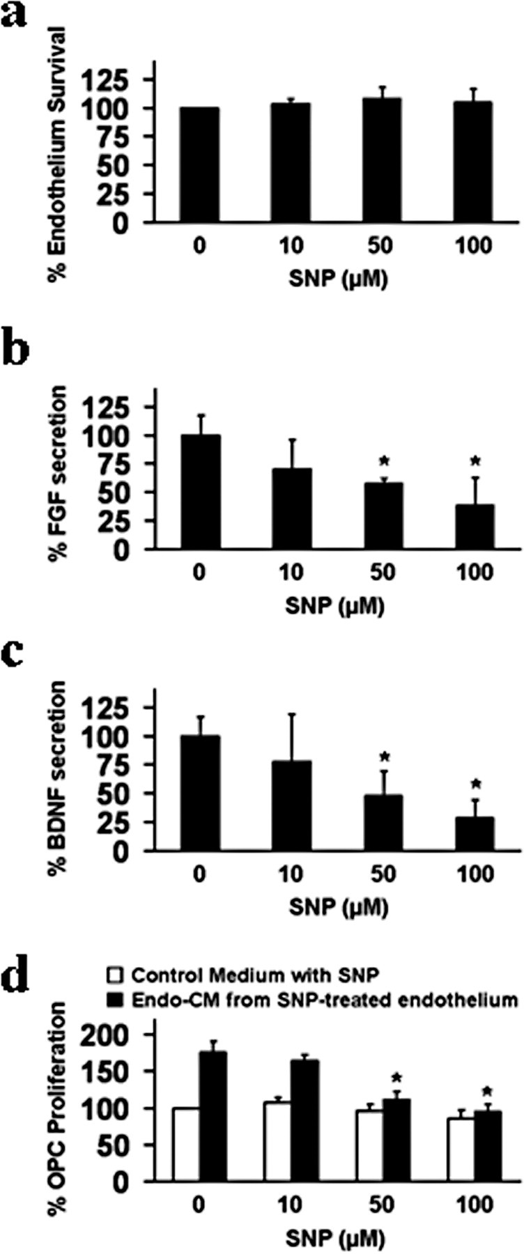
a, Sodium nitroprusside (SNP, 10–100 μm , exposure for 24 h) was not directly cytotoxic to primary human brain endothelial cells. b, c, But at these nonlethal concentrations, SNP significantly downregulated FGF and BDNF production (ELISA) by human brain endothelial cells. d, Conditioned media from primary human brain endothelial cells injured by nonlethal SNP no longer promoted OPC proliferation. Control OPC cultures were treated with empty media with matching levels of SNP. *p < 0.05.
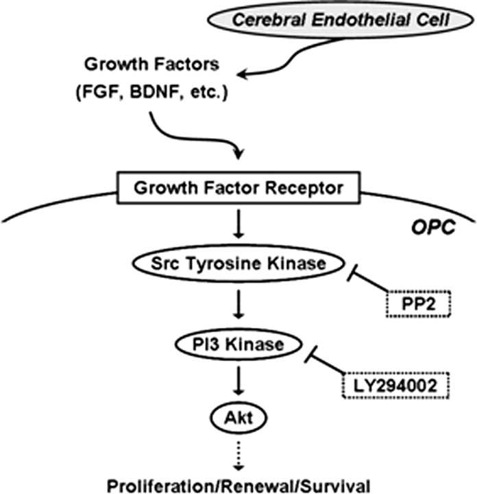
A proposed oligovascular niche; cerebral endothelial cells release trophic factors that activate Src, PI3 kinase, and Akt signaling to promote OPC proliferation. Oxidative stress or blockade of Src and Akt signaling prevents endothelial trophic factors from supporting OPCs.
References
Publication types
MeSH terms
Grants and funding
LinkOut - more resources
Full Text Sources
Medical
Miscellaneous