Comparative Study
. 2009 Apr 8;29(14):4381-91.
doi: 10.1523/JNEUROSCI.0377-09.2009.

Is that within reach? fMRI reveals that the human superior parieto-occipital cortex encodes objects reachable by the hand

Affiliations
Comparative Study

Is that within reach? fMRI reveals that the human superior parieto-occipital cortex encodes objects reachable by the hand

Jason P Gallivan et al. J Neurosci. .

Abstract

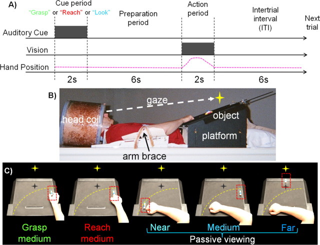
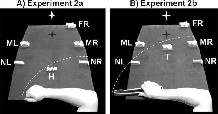
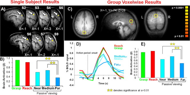
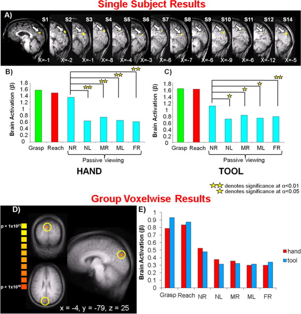
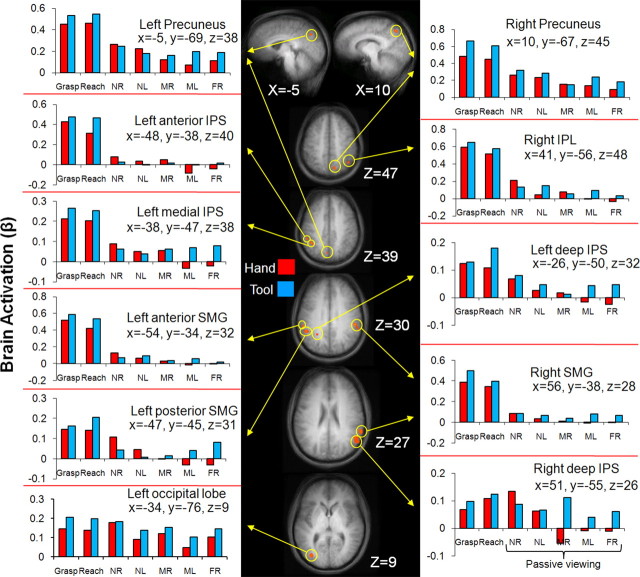
Macaque neurophysiology and human neuropsychology results suggest that parietal cortex encodes a unique representation of space within reach of the arm. Here, we used slow event-related functional magnetic resonance imaging (fMRI) to investigate whether human brain areas involved in reaching are more activated by objects within reach versus beyond reach. In experiment 1, graspable objects were placed at three possible locations on a platform: two reachable locations and one beyond reach. On some trials, participants reached to touch or grasp objects at the reachable location; on other trials participants passively viewed objects at one of the three locations. A reach-related area in the superior parieto-occipital cortex (SPOC) was more activated for targets within reach than beyond. In experiment 2, we investigated whether this SPOC response occurred when visual and motor confounds were controlled and whether it was modulated when a tool extended the effective range of the arm. On some trials, participants performed grasping and reaching actions to a reachable object location using either the hand alone or a tool; on other trials, participants passively viewed reachable and unreachable object locations. SPOC was significantly more active for passively viewed objects within reach of the hand versus beyond reach, regardless of whether or not a tool was available. Interestingly, these findings suggest that neural responses within brain areas coding actions (such as SPOC for reaching) may reflect automatic processing of motor affordances (such as reachability with the hand).


Figures

Figure 1.
Figure 1.
Experimental paradigm, setup, and conditions for experiment 1. A, Timing of one event-related trial. Participants begin trial by maintaining fixation. Participants receive an auditory instruction (gray square) to perform either a hand action (“grasp” or “reach”) or passive viewing (“look”) before presentation of the object stimuli. This is followed by a preparation period where participants prepare to perform the instruction. The action period is denoted by the illumination of the object (gray square). This is the cue for participants to perform the auditory instruction (pink dashed line represents the arm movement for grasp and reach trials only). This is then followed by an intertrial interval (ITI) until onset of the next trial. B, Picture of participant setup from side view. Participants' heads are tilted to permit direct viewing of objects on the platform. C, Experimental conditions as shown from participants' point of view. The yellow star with dark shadow represents the fixation LED and its location in depth (no shadow was actually visible). The yellow dashed line represents the extent of the participant's reaching space within the setup. The red dashed line denotes the location of the target object on the platform.
Figure 2.
Figure 2.
Experimental setup and conditions for experiment 2. On any given trial only one object would appear at any given location. A, Conditions for experiment 2a with the hand from the participant's point of view. During action trials, participants grasped and reached objects at location H with their hand while on passive viewing trials participants viewed an object at one of five locations: NR (near right), NL (near left), MR (middle right), ML (middle left), and FR (far right). B, Conditions for experiment 2b with the tool from the participant's point of view. Participants grasped and reached to objects at location T using the tool during action trials and on passive viewing trials, passively viewed objects in the same locations as in A. The white star with dark shadow represents the fixation LED and its location in depth (no shadow was actually visible). The white dashed line represents the extent of the participant's reaching space within the setup. Note that objects in medium and far locations are physically larger in size to provide a retinal size equivalent to that of the near objects.
Figure 3.
Figure 3.
Single-subject and group voxelwise results for experiment 1. A, Overlay of fMRI region-of-interest activation for the passive viewing of reachable versus unreachable objects [(Nearpv + Mediumpv) vs 2* Farpv] on 7 of 10 individual participants' sagittal slices (accompanying each slice is the X Talairach coordinate for the region of interest denoted by the arrow). Dashed white line corresponds to the parieto-occipital sulcus. B, Averaged β weights from the selected areas in A. C, Overlay of fMRI activation for reachable versus unreachable objects [(Nearpv vs Farpv) and (Mediumpv vs Farpv)] on a group averaged anatomical (using RFX analysis). Region defined by yellow circle denotes SPOC. D, % BOLD signal change from the selected region in C. E, β Weight activation from the selected region in C.
Figure 4.
Figure 4.
Single-subject and group voxelwise results for experiment 2. A, Overlay of fMRI region-of-interest activation for the passive viewing of reachable versus unreachable objects with respect to the hand [4*NRpv vs (NLpv + MRpv + MLpv + FRpv)] on 13 of 14 individual participants' sagittal slices (accompanying each slice is the X Talairach coordinate for the region of interest denoted by the arrow). Dashed white line corresponds to the parieto-occipital sulcus. B, Averaged β weights from the selected areas in A for functional runs with the hand. C, Averaged β weights from selected areas in A for functional runs with the tool. D, Overlay of fMRI activation for reachable versus unreachable objects [4*NRpv vs (NLpv + MRpv + MLpv + FRpv)] on a group averaged anatomical (using FFX analysis, cluster threshold corrected, 380 mm3). Region defined by yellow circle denotes SPOC. Accompanying the region of interest are its Talairach coordinates. E, β Weight activation from the selected region in D for functional runs with the hand and tool. Note that for both the hand and tool runs, post hoc comparisons confirm that NRpv is statistically higher than each of the nonreachable object locations (NLpv, MRpv, MLpv, and FRpv). NR, Near right; NL, near left; MR, middle right; ML, middle left; FR, far right.
Figure 5.
Figure 5.
Areas activated by a group voxelwise contrast for the passive viewing of reachable versus unreachable objects with respect to the hand [NRpv vs (NLpv + MRpv + MLpv + FRpv)] overlayed on group averaged anatomical slices (using RFX analysis). Located near each anatomical slice is its relevant Talairach coordinate. fMRI activation is shown in orange at p < 0.001 with its accompanying β weight activation denoted by yellow arrows. Note that the scale of brain activation changes across brain areas. NR, Near right; NL, near left; MR, middle right; ML, middle left; FR, far right.

References

    1. Andersen RA. Neural mechanisms of visual motion perception in primates. Neuron. 1997;18:865–872. - PubMed
    1. Andersen RA, Buneo CA. Intentional maps in posterior parietal cortex. Ann Rev Neurosci. 2002;25:189–220. - PubMed
    1. Andersen RA, Snyder LH, Bradley DC, Xing J. Multimodal representation of space in the posterior parietal cortex and its use in planning movements. Ann Rev Neurosci. 1997;20:303–330. - PubMed
    1. Astafiev SV, Shulman GL, Stanley CM, Snyder AZ, Van Essen DC, Corbetta M. Functional organization of human intraparietal and frontal cortex for attending, looking, and pointing. J Neurosci. 2003;23:4689–4699. - PMC - PubMed
    1. Battaglini PP, Muzur A, Galletti C, Skrap M, Brovelli A, Fattori P. Effects of lesions to area V6A in monkeys. Exp Brain Res. 2002;144:419–422. - PubMed

Publication types