Comparative Study
. 2009 Apr 8;29(14):4408-19.
doi: 10.1523/JNEUROSCI.6003-08.2009.

Neural stem and progenitor cells retain their potential for proliferation and differentiation into functional neurons despite lower number in aged brain

Affiliations
Comparative Study

Neural stem and progenitor cells retain their potential for proliferation and differentiation into functional neurons despite lower number in aged brain

Henrik Ahlenius et al. J Neurosci. .

Abstract

Neurogenesis in the subventricular zone (SVZ), which gives rise to new neurons in the olfactory bulb, continues throughout life but declines with increasing age. Little is known about how aging affects the intrinsic properties of the neural stem and progenitor cells (NSCs) in SVZ and the functional characteristics of their neuronal progeny. Here, we have compared the properties of NSCs isolated from embryonic lateral ganglionic eminence and adult and aged SVZ in mice using in vivo and in vitro systems, analyzed their gene expression profile, and studied their electrophysiological characteristics before and after differentiation into neurons. We show a loss of NSCs in SVZ from aged mice accompanied by reduced expression of genes for NSC markers, developmentally important transcription factors, and neurogenic factors. However, when isolated in vitro, the NSCs from SVZ of aged animals have capacity for proliferation and multilineage differentiation, including production of functional neurons, similar to that of NSCs in adult mice, albeit with lower efficacy. These properties are of major importance when considering therapeutic applications of neuronal replacement from endogenous NSCs in the injured, aged brain.


Figures

Figure 1.
Figure 1.
Aging causes severe reduction of in vivo neurogenesis and loss of NSCs in SVZ. A–G, Proliferation as assessed by number of p-H3+ (A) and BrdU+ (B) cells, neuroblast formation (C) as assessed by Dcx+ cells, number of NSCs as assessed by number of nestin–GFP+ cells (D), and number of cells expressing the putative markers Sox2 (E) and Gsh2 (F), and proliferative activity in NSCs and neuroblasts as assessed by the percentage BrdU+/Sox2+ and BrdU+/Dcx+ cells of the total number of Sox2+ and Dcx+ (G) cells, respectively, in adult and aged SVZ. Means ± SEM; n = 6 and 4 (A–C and E, F), n = 6 and 3 (D), and n = 5 and 6 (G) for adult and aged, respectively. *p < 0.05, one-way ANOVA with Fisher's post hoc test. Scale bars: A–C, 100 μm; D, E, 30 μm.
Figure 2.
Figure 2.
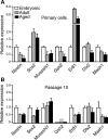
Aging causes reduction of NSC and neurogenic factor gene expression in SVZ stem/progenitor cells. A, B, Gene expression levels for stem/progenitor cell markers (nestin, Sox2, Musashi1), developmentally important transcription factors (Gsh2, Er81), and neurogenic factors (Dlx2, Mash1) measured using quantitative PCR in primary (A) or expanded (B) nestin–GFP+ cells sorted from embryonic LGE, and adult and aged SVZ of nestin–GFP mice. Data are presented as relative expression normalized to GAPDH levels and compared with embryonic LGE levels (set at 1). Means from two to four individual experiments. ND, Not detected.
Figure 3.
Figure 3.
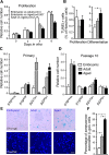
Decreased neurosphere formation in aged SVZ is normalized after enrichment for NSCs. A, B, Yield of primary, secondary, and passage 10 neurospheres from nonsorted cells (A) and sorted GFP+ cells (B), derived from embryonic LGE, and adult and aged SVZ in wild-type (A) and nestin–GFP mice (B). Means ± SEM (n = 5). *p < 0.05, one-way ANOVA with Fisher's post hoc test. Nr, Number.
Figure 4.
Figure 4.
Aging causes reduction of NSC proliferation and survival during differentiation in vitro. Neurosphere cells were grown under proliferative and differentiating conditions. A, Relative number of cells assessed by MTT test. Proliferation was significantly higher in embryonic compared with adult NSCs and in adult compared with aged NSCs at 0, 2, 4, and 6 DIV. Means ± SEM; *p < 0.05, repeated-measures ANOVA. Four independent cultures were used for quantification. B, Survival assessed by numbers of TUNEL+ apoptotic cells under proliferative (at 4 DIV) or differentiating conditions (at 7 DIV). Means ± SEM. Four independent cultures were used for quantification. C, D, Expression of genes involved in senescence in primary (C) and expanded (D) Nestin–GFP+ sorted cells. E, F, Photomicrograph (E) and percentage (F) of neurosphere cells, from the different age groups, expressing SA-β-gal. Mean ± SEM. Four independent cultures were used for quantification. *p < 0.05, one-way ANOVA with Fisher's post hoc test. Scale bar, 30 μm. ND, Not detected.
Figure 5.
Figure 5.
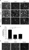
Multilineage differentiation of embryonic, adult, and aged NSCs. A, Photomicrographs of cells with morphological and immunocytochemical characteristics of neurons (β-III-tubulin+), astrocytes (GFAP+), and oligodendrocytes (CNPase+) generated after differentiation (7 DIV) of neurospheres derived from embryonic LGE and adult and aged SVZ. B, Percentage of β-III-tubulin+ cells out of all cells formed from embryonic LGE and adult and aged SVZ. Mean ± SEM; *p < 0.05, one-way ANOVA with Fisher's post hoc test. Four independent cultures were used for quantification. C, Photomicrograph showing a glutamate+ cell and a GABA+ cell derived from aged NSCs. The GABA+ cell was a functional neuron as assessed by patch-clamp recordings of the same cell (indicated by biocytin labeling). Scale bar, 20 μm.
Figure 6.
Figure 6.
Active membrane properties of embryonic, adult, and aged NSCs. A, Responses to depolarizing or hyperpolarizing current injections in embryonic, adult, and aged proliferating (top) and differentiated (bottom) NSCs. Currents were injected for 500 ms in 20 pA increments every 5 s. Insets show microscopic images of corresponding cells with arrowheads depicting the recorded cells. Complete blockade of discharge responses to depolarizing current injections of 10 pA in differentiated cells by 1 μm TTX is also shown. Calibration: 20 mV, 200 ms. n = 16, 13, 13 (top), and 23, 22, and 11 (bottom) for embryonic, adult, and aged, respectively. B, Percentage of neurons responding to membrane depolarization with generation of immature, mature, or no AP embryonic (n = 26), adult (n = 38), and aged group (n = 42). C, Expression of Na+ currents in neurons differentiated from embryonic, adult, and aged NSCs induced by voltage steps from −70 to 0 mV in the absence and presence of 1 μm TTX. Calibration: 200 pA, 5 ms. n = 6 for embryonic, 7 for adult, and 7 for aged.
Figure 7.
Figure 7.
Expression of K+ currents in proliferating cells. A, Total K+ current (Ik(DR+A); left), non-inactivating delayed-rectified current (IK(DR); middle), and a fast-inactivating A-type K+ current (IK(A); right) obtained by voltage steps from −70 to +60 mV after a hyperpolarizing prepulse to −110 mV in proliferating embryonic, adult, and aged NSCs. IK(DR) was separated from total IK(A) using a depolarizing prepulse to −40 mV. Calibration: 1 nA, 50 ms. B, K+ current density at +60 mV. C, D, Mean ± SEM. Mean proportion IK(A) (C), and IK(DR) (D) out of total current plotted against the applied voltage steps for embryonic, adult, and aged NSCs, respectively. In C, comparison with repeated-measures ANOVA. n = 19, 20, 21 in embryonic, adult, and aged, respectively.
Figure 8.
Figure 8.
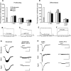
K+ channel sensitivity to TEA and 4AP in embryonic, adult, and aged NSCs. A, Percentage of the current left unblocked by 20 mm TEA, 2 mm 4AP, or 20 mm TEA and 2 mm 4AP in proliferating embryonic, adult, and aged NSCs. Means ± SEM; n = 11, 10, 7 and 9, 9, 7 and 17, 17, 13 in embryonic, adult, and aged, respectively. B, C, Representative traces of outward K+ currents activated by a depolarizing voltage step from the resting membrane potential to +40 mV in proliferating NSCs. The currents are blocked by 20 mm TEA (B) or 2 mm 4AP (C). Calibration: 500 pA, 25 ms. D, Percentage of the current left unblocked by 20 mm TEA, 2 mm 4AP, or 20 mm TEA and 2 mm 4AP in neurons differentiated from embryonic, adult, and aged NSCs. Means ± SEM; n = 5, 5, 5 and 6, 5, 7 and 8, 10, 11 in embryonic, adult, and aged, respectively. E, F, Representative traces of outward K+ currents activated by a depolarizing voltage step from the resting membrane potential to +40 mV in neurons differentiated from aged NSCs. The currents are blocked by 20 mm TEA (E) or 2 mm 4AP (F). Calibration: 250 pA, 25 ms. G, Effect of 100 μm glutamate (left) or glutamate together with 10 μm NBQX and 50 μm d-AP5 (right) on neurons differentiated from embryonic, adult, and aged NSCs. Calibration: for embryonic and aged, 100 pA, 50 ms; for adult cells, 20 pA, 50 ms. n = 6, 8, and 6 in embryonic, adult, and aged, respectively. H, The effect of 100 μm GABA (left) or 100 μm GABA together with 10 μm PTX (right) on neurons differentiated from embryonic, adult, and aged NSCs. Calibration: for embryonic and aged, 500 pA, 50 ms; for adult cells, 40 pA and 50 ms. n = 8, 7, and 5 in embryonic, adult, and aged, respectively.

References

    1. Bailey KJ, Maslov AY, Pruitt SC. Accumulation of mutations and somatic selection in aging neural stem/progenitor cells. Aging Cell. 2004;3:391–397. - PubMed
    1. Barish ME. Differentiation of voltage-gated potassium current and modulation of excitability in cultured amphibian spinal neurones. J Physiol. 1986;375:229–250. - PMC - PubMed
    1. Bortner CD, Hughes FM, Jr, Cidlowski JA. A primary role for K+ and Na+ efflux in the activation of apoptosis. J Biol Chem. 1997;272:32436–32442. - PubMed
    1. Brown JP, Couillard-Després S, Cooper-Kuhn CM, Winkler J, Aigner L, Kuhn HG. Transient expression of doublecortin during adult neurogenesis. J Comp Neurol. 2003;467:1–10. - PubMed
    1. Cameron HA, McKay RD. Restoring production of hippocampal neurons in old age. Nat Neurosci. 1999;2:894–897. - PubMed

Publication types