Comparative Study
. 2009 Apr 8;29(14):4461-70.
doi: 10.1523/JNEUROSCI.0296-09.2009.

Conditional inactivation of androgen receptor gene in the nervous system: effects on male behavioral and neuroendocrine responses

Affiliations
Comparative Study

Conditional inactivation of androgen receptor gene in the nervous system: effects on male behavioral and neuroendocrine responses

Kalina Raskin et al. J Neurosci. .

Abstract

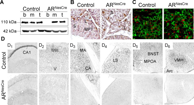
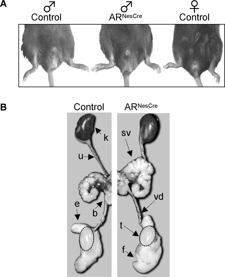
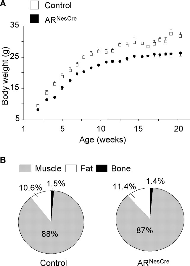
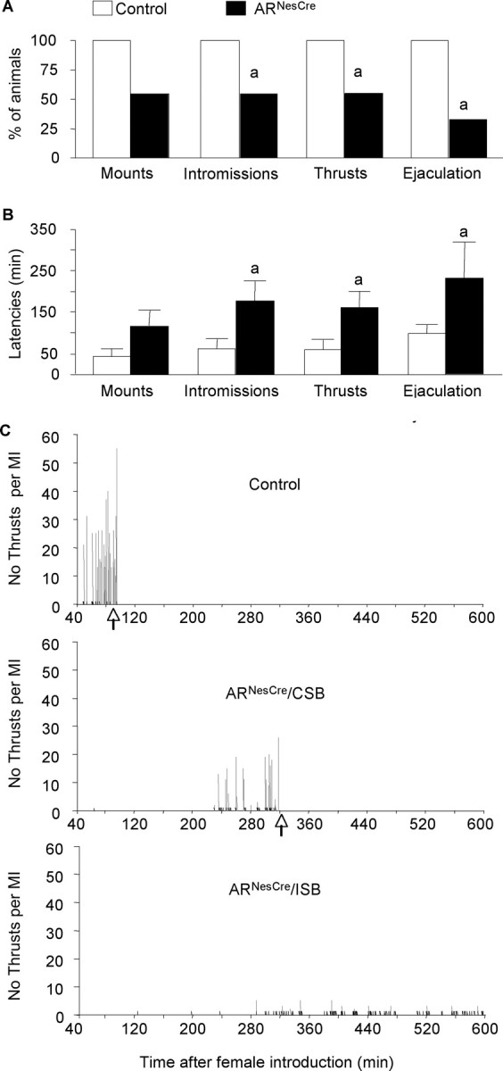
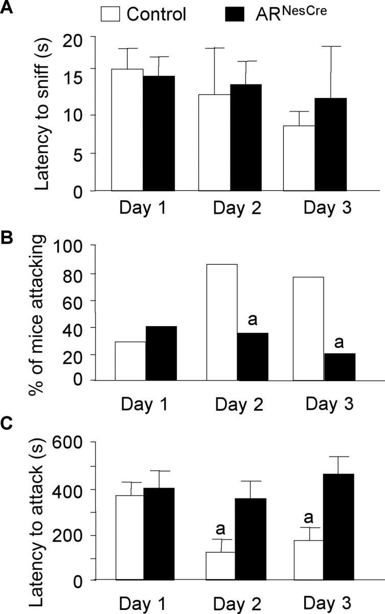
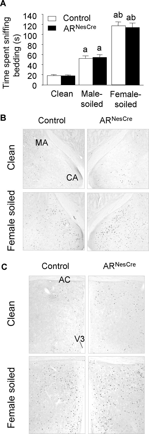
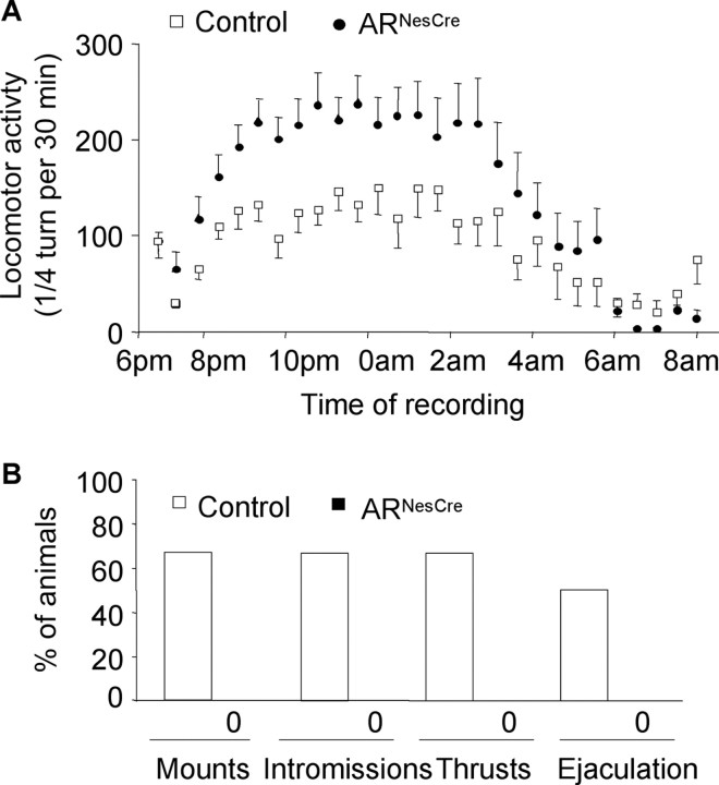
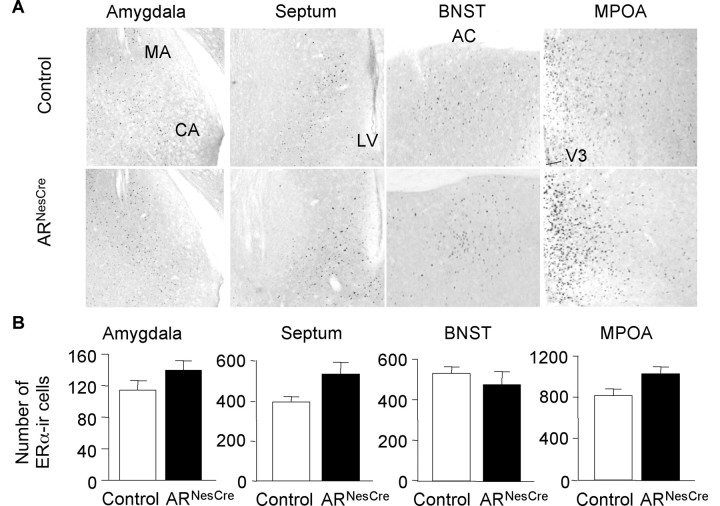
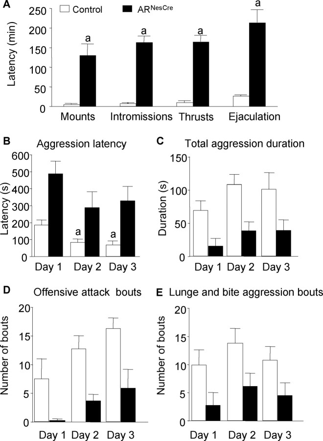
Testosterone (T) profoundly influences central sexual differentiation and functions. In the brain, T signals either directly through androgen receptor (AR) or indirectly through estrogen receptor (ER) following aromatization into E2 (17-beta-estradiol). As T, through AR, also controls peripheral male sexual differentiation, the relative contribution of central AR in T-mediated regulation of behavioral and neuroendocrine responses still remains unclear. To address this question, we generated, by using Cre-loxP technology, mice selectively lacking AR expression in the nervous system. The mutant male urogenital tract was normally developed, and mice were able to produce offspring. Nonetheless, sexual motivation and performance as well as aggressive behaviors were affected. Only a low percentage of males displayed a complete sexual behavior and offensive attacks. The latency to show masculine behaviors was increased and copulation length prolonged. Erectile activity during mating was also altered. These alterations occurred despite increased levels of T and its metabolites, and an unaffected number of ERalpha-immunoreactive cells. Olfactory preference and neuronal activation, mapped by Fos immunoreactivity, following exposure to estrus female-soiled bedding were also normal. At comparable T levels, greater differences in masculine behaviors were observed between gonadectomized control and mutant males. AR invalidation in the nervous system also disrupted the somatotropic axis since mutant males exhibited growth retardation and decreased serum levels of insulin-like growth factor I. Our findings show that central AR is required in T-induced regulation of male-typical behaviors and gonadotrope and somatotropic axes. This genetic model offers a unique opportunity in the understanding of AR's role in cerebral functions of T.


Figures

Figure 1.
Figure 1.
AR gene disruption is specific to the nervous system in ARNesCre mice. A, Western blotting of the 110 kDa AR and 42 kDa actin proteins from brain (b), skeletal muscle (m), and testicular (t) extracts of control and ARNesCre mice. B, Immunostaining of AR protein in testicular sections. AR staining was found in Leydig (L), Sertoli (S), and myoid peritubular (MPT) cells of control and ARNesCre mice. C, Immunofluorescent detection of AR protein (red) and LHβ subunit (green) in pituitary sections. D, Immunostaining of AR protein in coronal brain sections. In control brain, AR protein was detected in the CA1 region of hippocampus (D1), II/III/V layers of the sensorimotor cortex (D2), MA and cortical amygdala (CA) (D3), lateral septum (LS) (D4), BNST and different hypothalamic regions, including the MPOA (D5), and arcuate nucleus (Arc) and ventromedial hypothalamus (VMH) (D6). The corresponding ARNesCre sections show no specific AR immunostaining.
Figure 2.
Figure 2.
Male phenotype of genetic (XY) ARNesCre mice. A, External sexual development of ARNesCre males was compared with control male and female littermates at 3–4 months of age. Anogenital distance was similar between control (10.8 ± 0.6 mm) and mutant (11.1 ± 1.2 mm) males (n = 7–10 per genotype). B, Urogenital tract of control and ARNesCre males. k, Kidney; sv, seminal vesicle; vd, vas deferens; t, testis; b, bladder; f, fat tissue; e, epididymis; u, ureter.
Figure 3.
Figure 3.
Growth of ARNesCre mice. A, Control and ARNesCre males from the same litters (n = 15–19 per genotype) were weekly weighed. The growth curves are significantly different (p < 0.05). B, Body composition of live adult (3 months old) males (n = 5 per genotype) was analyzed by using a Piximus densitometer (Lunar Corporation). Muscle, fat, and bone masses are given as percentages of total body weight.
Figure 4.
Figure 4.
Male sexual behavior of control and ARNesCre mice measured in a 10 h test. A, Percentage of males showing mounts, mounts with intromissions, thrusts with intromissions, and ejaculation. B, Latency to the first mount (without intromission), intromission, thrust, and ejaculation for mice that displayed complete sexual behavior (n = 9–11 per genotype; ap < 0.05 vs control mice). C, Representative time courses of male sexual behavior of control and mutant males showing complete (CSB) or incomplete (ISB) sexual behavior. The occurrences of mounts without intromissions (black lines), mounts with intromissions (MI) (gray lines), and ejaculation (arrows) within the 600 min of the test are indicated. The total number of thrusts for each MI is represented on the y-axis.
Figure 5.
Figure 5.
Aggressive behavior of mice in the resident–intruder paradigm over 3 consecutive days. A, Latency to anogenital chemoinvestigation of intruders with no significant effect of genotype (F(1,40) = 0.62, p = 0.44). B, Percentage of males showing aggressive bouts on days 1, 2, and 3 (ap < 0.05 vs control mice). C, Latency to attack intruder mice. For residents that did not show aggressive behavior on days 2 and 3 of the test, the latency was 600 and 1200 s, respectively. There was a significant effect of genotype (F(1,24) = 8.55, p < 0.05). n = 20 per genotype; post hoc analysis showed significant decreased latency for control mice to attack at days 2 or 3 versus day 1 (ap < 0.05).
Figure 6.
Figure 6.
Olfactory preference and neuronal activation. A, Time spent chemoinvestigating clean, male-soiled, and female-soiled-bedding by control and ARNesCre mice (n = 20 per genotype). A significant effect of bedding (F(2,76) = 143.58, p < 0.0001) but not of genotype was found; post hoc analysis showed differences in the time spent sniffing the three beddings (ap < 0.001 vs clean bedding; bp < 0.001 vs male-soiled bedding). B, C, Representative c-fos immunostaining in the medial amygdala (B) and medial preoptic area (C) of control and ARNesCre males exposed to clean or female-soiled beddings for 1 h. CA, Cortical amygdala; AC, anterior commissure; V3, third ventricle.
Figure 7.
Figure 7.
Locomotor activity and effect of DHT treatment on sexual behavior of control and ARNesCre males. A, Time course of an overnight 14 h recording of locomotor activity. The test started 1 h before dark phase (6:00 P.M.) and ended 1 h after lights on (8:00 A.M.). n = 8–13 per genotype. B, Sexual behavior of gonadectomized and DHT-treated control and ARNesCre males in the 10 h test (n = 6 per genotype). Sexual behavior length for control mice was similar to that of intact control males (57 ± 15 min).
Figure 8.
Figure 8.
Quantification of ERα-IR neurons in chemoresponsive brain areas of control and ARNesCre mice. A, Representative anti-ERα immunostaining in amygdala, septum, BNST, and MPOA. CA, Cortical amygdala; LV, lateral ventricle; AC, anterior commissure; V3, third ventricle. B, Quantitative data (n = 3–6 per genotype) expressed as ERα-positive cells per unit area.
Figure 9.
Figure 9.
Male sexual and aggressive behaviors of gonadectomized and T-treated mice. A, Latency to the first mount, mount with intromission, thrust, and ejaculation in the 10 h test (n = 6 per genotype; ap < 0.05 vs control mice). B, Latency to the first aggressive behavioral act in the 10 min resident–intruder paradigm with a significant effect of genotype (F(1,28) = 10.99, p = 0.0051); post hoc analysis showed a significantly decreased latency for control mice to attack at days 2 or 3 versus day 1 (ap < 0.05). C, The total aggression duration was decreased in mutant mice (F(1,28) = 20.13, p = 0.0005). D, The number of offensive attacks was lower in ARNesCre mice (F(1,28) = 16.71, p = 0.0011). E, The number of lunges and bites was reduced for ARNesCre males (F(1,28) = 20.13, p = 0.0005). n = 8 per genotype.

References

    1. Agmo A. Male rat sexual behavior. Brain Res Brain Res Protoc. 1997;1:203–209. - PubMed
    1. Arteaga-Silva M, Rodríguez-Dorantes M, Baig S, Morales-Montor J. Effects of castration and hormone replacement on male sexual behavior and pattern of expression in the brain of sex-steroid receptors in BALB/c AnN mice. Comp Biochem Physiol A Mol Integr Physiol. 2007;147:607–615. - PubMed
    1. Baker J, Hardy MP, Zhou J, Bondy C, Lupu F, Bellvé AR, Efstratiadis A. Effects of an Igf1 gene null mutation on mouse reproduction. Mol Endocrinol. 1996;10:903–918. - PubMed
    1. Bakker J, Honda S, Harada N, Balthazart J. Sexual partner preference requires a functional aromatase (cyp19) gene in male mice. Horm Behav. 2002;42:158–171. - PubMed
    1. Bodo C, Rissman EF. Androgen receptor is essential for sexual differentiation of responses to olfactory cues in mice. Eur J Neurosci. 2007;25:2182–2190. - PubMed

Publication types

Substances