Comparative Study
. 2009 Apr 8;29(14):4664-74.
doi: 10.1523/JNEUROSCI.5502-08.2009.

Activity profiles of cholinergic and intermingled GABAergic and putative glutamatergic neurons in the pontomesencephalic tegmentum of urethane-anesthetized rats

Affiliations
Comparative Study

Activity profiles of cholinergic and intermingled GABAergic and putative glutamatergic neurons in the pontomesencephalic tegmentum of urethane-anesthetized rats

Soufiane Boucetta et al. J Neurosci. .

Abstract

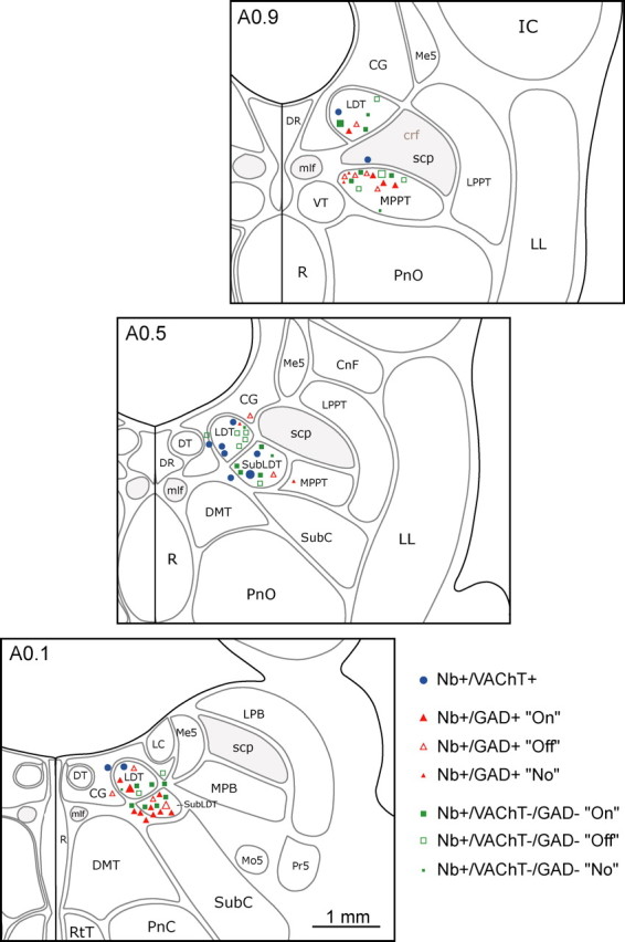
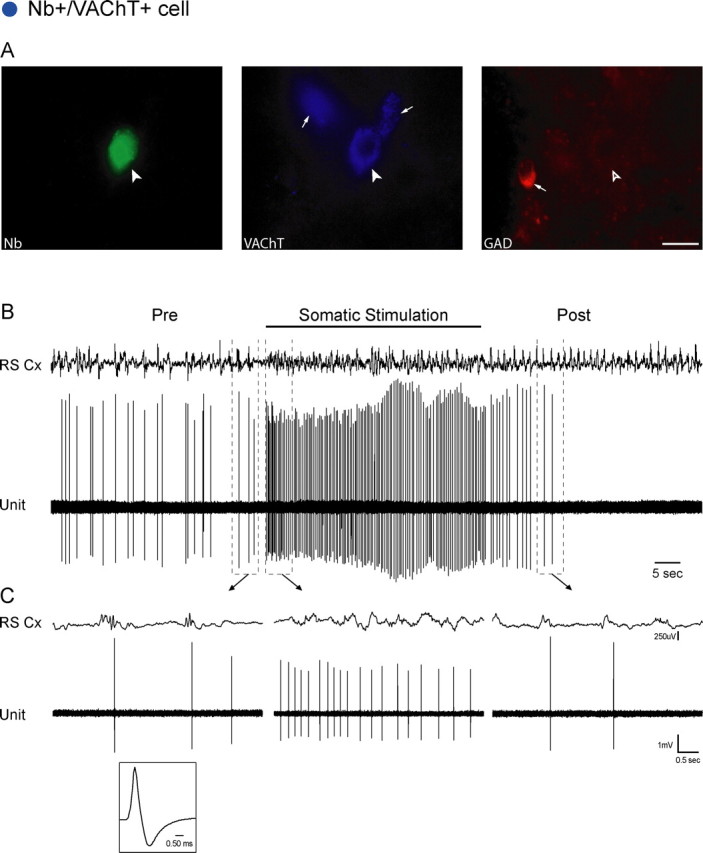
Cholinergic neurons in the pontomesencephalic tegmentum form part of the ascending activating system and are thought to participate in stimulating cortical activation. Yet in the laterodorsal tegmental and pedunculopontine tegmental nuclei (LDT and PPT), they lie intermingled with GABAergic and glutamatergic neurons, which could also modulate cortical activity and sleep-wake state. To characterize the discharge of these cell types in relation to cortical activity, we recorded neurons in urethane-anesthetized rats during spontaneous slow wave and somatosensory evoked fast electroencephalographic (EEG) activity, then labeled the cells by juxtacellular technique with Neurobiotin (Nb) and dual-immunostained them for vesicular acetylcholine transporter (VAChT) and glutamic acid decarboxylase (GAD). All cholinergic cells discharged minimally during prestimulation (approximately 0.5 Hz) and moderately in a tonic manner (approximately 4 Hz) during stimulation. Being heterogeneous, some GABAergic, called "On," cells (approximately 48%) increased their discharge (from approximately 4 to 7 Hz), whereas others, called "Off" cells (approximately 38%), decreased or ceased firing during stimulation. Similarly, some noncholinergic/non-GABAergic On cells increased (from approximately 2 to 6 Hz, approximately 49%), whereas other Off cells decreased firing ( approximately 35%) during stimulation. Putative glutamatergic On together with GABAergic On neurons could thus act in parallel with cholinergic cells to stimulate cortical activation. Possibly influenced by cholinergic On and glutamatergic Off cells, whose change in discharge precedes theirs, the GABAergic Off cells could oppose neighboring neurons such as noradrenergic cells, which discharge during waking and cease firing during sleep. By concerted activity, these heterogeneous cell groups can modulate cortical activity and behavioral state across the sleep-waking cycle.


Figures

Figure 1.
Figure 1.
Distribution of recorded cells in the mesopontine tegmentum. The recorded, Nb-labeled cells were immunohistochemically identified as cholinergic, using VAChT (Nb+/VAChT+, blue circles), as GABAergic, using GAD (Nb+/GAD+, red triangles), or as noncholinergic/non-GABAergic (Nb+/VAChT−/GAD−, green squares). GABAergic and noncholinergic/non-GABAergic cells were further distinguished according to their response as On (filled symbols), Off (open symbols), or No (small filled symbols) response to somatic stimulation. Cells are mapped onto appropriate levels (anterior, A0.9, A0.5, or A0.1 mm to interaural zero) through the LDT/SubLDT/MPPT cholinergic cell area. Recordings and images are presented for representative cells (largest symbols) of the cholinergic (Fig. 2), GABAergic On and Off (Figs. 3, 4), and noncholinergic/non-GABAergic On and Off cells (Figs. 5, 6). CG, Central gray; CnF, cuneiform nucleus; crf, central reticular fasciculus; DMT, dorsomedial tegmental area; DR, dorsal raphe nucleus; DT, dorsal tegmental nucleus; IC, inferior colliculus; LC, locus ceruleus; LL, lateral lemniscus; LPB, lateral parabrachial nucleus; LPPT, lateral pedunculopontine tegmental nucleus; Me5, mesencephalic trigeminal nucleus; mlf, medial longitudinal fasciculus; Mo5, motor trigeminal nucleus; MPB, medial parabrachial nucleus; PnC, pontine reticular nucleus, caudal part; PnO, pontine reticular nucleus, oral part; Pr5, principal sensory trigeminal nucleus; R, raphe nuclei; RtT, reticulotegmental nucleus of the thalamus; scp, superior cerebellar peduncle; SubC, subceruleus; VT, ventral tegmental nucleus.
Figure 2.
Figure 2.
Nb+/VAChT+ cell. A, The Nb-labeled cell (filled arrowhead, #ABS80a) was positively immunostained for VAChT (filled arrowhead) and negatively for GAD (open arrowhead), while near other VAChT+ and GAD+ cells in the region (small arrows). Scale bar, 20 μm. The cell was located in the SubLDT (largest blue circle) (Fig. 1, A0.5). B, C, The unit discharged at an average low rate (0.70 Hz) in association with spontaneous irregular slow-wave activity on the EEG of the retrosplenial cortex (RS Cx) in the period preceding stimulation (Pre). It increased its rate markedly (to 3.10 Hz) and fired tonically during somatic stimulation in association with faster activity on the EEG. (See also supplemental Figs. 2, 3, available at www.jneurosci.org as supplemental material.) Note that the increase in unit discharge preceded the change in EEG activity (by ∼180 ms). After stimulation (Post), the unit initially decreased then ceased firing as the EEG returned to irregular slow-wave activity. Traces in B (enclosed by dashed lines) are expanded in C (arrows). The unit had a relatively long duration spike (shown in C, 0.76 and 2.55 ms at first and second zero crossings) (supplemental Fig. 2, available at www.jneurosci.org as supplemental material).
Figure 3.
Figure 3.
Nb+/GAD+ On cell. A, Nb-labeled cell (filled arrowhead, #ABS78b) was negatively immunostained for VAChT (open arrowhead) and positively so for GAD (filled arrowhead), while located near other VAChT+ and GAD+ cells in the region (small arrows). Scale bar, 20 μm. The cell was situated in the LDT (largest red filled triangle) (Fig. 1, A0.1). B, C, The unit discharged at a low average rate (0.46 Hz) in association with irregular slow-wave activity on the EEG of the retrosplenial cortex (RS Cx) before stimulation (Pre). It increased its firing markedly (to 11.49 Hz) and fired tonically in association with faster cortical activity riding upon rhythmic slow (theta-like) activity during somatic stimulation. Note that the increase in unit discharge followed the change in EEG activity (by ∼1.22 s). After stimulation (Post), the unit decreased its rate of firing to return to prestimulation levels in similar association with irregular slow-wave activity. Traces in B (enclosed by dashed lines) are expanded in C (arrows). The unit had a relatively short duration spike (shown in C, 0.44 and 1.21 ms at first and second zero crossings).
Figure 4.
Figure 4.
Nb+/GAD+ Off cell. A, Nb-labeled cell (filled arrowhead, #ABS102a) was negatively immunostained for VAChT (open arrowhead) and positively so for GAD (filled arrowhead), while located near other VAChT+ and GAD+ cells in the region (small arrows). Scale bar, 20 μm. The cell was situated in the SubLDT (largest red open triangle) (Fig. 1, A0.1). B, C, The unit discharged phasically at a moderate average rate (2.07 Hz) in association with irregular slow-wave activity on the EEG of the retrosplenial cortex (RS Cx) before stimulation (Pre). It ceased firing in association with faster cortical activity riding upon rhythmic slow (theta-like) activity during somatic stimulation. Note that the unit ceased firing following the change in EEG activity (by ∼1.31 s). After stimulation (Post), the unit recovered its baseline rate of firing in association with irregular slow-wave activity. Traces in B (enclosed by dashed lines) are expanded in C (arrows). The unit had a relatively short duration spike (shown in C, 0.35 and 0.93 ms at first and second zero crossings).
Figure 5.
Figure 5.
Nb+/VAChT−/GAD− On cell. A, Nb-labeled cell (filled arrowhead, #ABS87c) was negatively immunostained for VAChT (open arrowhead) and for GAD (open arrowhead), while located near other VAChT+ and GAD+ cells in the region (small arrows). Scale bar, 20 μm. The cell was situated in the LDT (largest green filled square) (Fig. 1, A0.9). B, C, The unit discharged irregularly at a low average rate (0.385 Hz) in association with irregular slow-wave activity on the EEG of the retrosplenial cortex (RS Cx) before stimulation (Pre). It increased its firing markedly (to 3.59 Hz) and fired tonically in association with faster cortical activity during somatic stimulation. Note that the increase in unit discharge preceded the change in EEG activity (by ∼210 ms). After stimulation (Post), the unit decreased its rate back to baseline levels in association with irregular slow-wave activity. Traces in B (enclosed by dashed lines) are expanded in C (arrows). The unit had a relatively short duration spike (shown in C, 0.36 and 0.98 ms at first and second zero crossings).
Figure 6.
Figure 6.
Nb+/VAChT−/GAD− Off cell. A, Nb-labeled cell (filled arrowhead, #ABS54) was negatively immunostained for VAChT (open arrowhead) and for GAD (open arrowhead), while located near other VAChT+ and GAD+ cells in the region (small arrows). Scale bar, 20 μm. The cell was situated in the MPPT (largest green open square) (Fig. 1, A0.9). B, C, The unit discharged at a fast average rate (13.28 Hz) in association with irregular slow-wave activity on the EEG of the retrosplenial cortex (RS Cx) before stimulation (Pre). It decreased its average discharge rate (to 4.20 Hz) in association with faster cortical activity during somatic stimulation. After stimulation (Post), the unit recovered its baseline rate in association with irregular slow-wave activity. Traces in B (enclosed by dashed lines) are expanded in C (arrows). The unit had a medium duration spike (shown in C, 0.49 and 1.34 ms at first and second zero crossings).

References

    1. Amatruda TT, 3rd, Black DA, McKenna TM, McCarley RW, Hobson JA. Sleep cycle control and cholinergic mechanisms: differential effects of carbachol injections at pontine brain stem sites. Brain Res. 1975;98:501–515. - PubMed
    1. Aston-Jones G, Bloom FE. Activity of norepinephrine-containing locus coeruleus neurons in behaving rats anticipates fluctuations in the sleep–waking cycle. J Neurosci. 1981;1:876–886. - PMC - PubMed
    1. Brischoux F, Mainville L, Jones BE. Muscarinic-2 and orexin-2 receptors on GABAergic and other neurons in the rat mesopontine tegmentum and their potential role in sleep-wake state control. J Comp Neurol. 2008;510:607–630. - PubMed
    1. Brown RE, McKenna JT, Winston S, Basheer R, Yanagawa Y, Thakkar MM, McCarley RW. Characterization of GABAergic neurons in rapid-eye-movement sleep controlling regions of the brainstem reticular formation in GAD67-green fluorescent protein knock-in mice. Eur J Neurosci. 2008;27:352–363. - PMC - PubMed
    1. Clement EA, Richard A, Thwaites M, Ailon J, Peters S, Dickson CT. Cyclic and sleep-like spontaneous alternations of brain state under urethane anaesthesia. PLoS ONE. 2008;3:e2004. - PMC - PubMed

Publication types

LinkOut - more resources