. 2009 Apr 28;106(17):6939-44.
doi: 10.1073/pnas.0900833106. Epub 2009 Apr 8.

A single amino acid change converts Aurora-A into Aurora-B-like kinase in terms of partner specificity and cellular function

Affiliations

A single amino acid change converts Aurora-A into Aurora-B-like kinase in terms of partner specificity and cellular function

Jingyan Fu et al. Proc Natl Acad Sci U S A. .

Abstract

Aurora kinase-A and -B are key regulators of the cell cycle and tumorigenesis. It has remained a mystery why these 2 Aurora kinases, although highly similar in protein sequence and structure, are distinct in subcellular localization and function. Here, we report the striking finding that a single amino acid residue is responsible for these differences. We replaced the Gly-198 of Aurora-A with the equivalent residue Asn-142 of Aurora-B and found that in HeLa cells, Aurora-A(G198N) was recruited to the inner centromere in metaphase and the midzone in anaphase, reminiscent of the Aurora-B localization. Moreover, Aurora-A(G198N) compensated for the loss of Aurora-B in chromosome misalignment and cell premature exit from mitosis. Furthermore, Aurora-A(G198N) formed a complex with the Aurora-B partners, INCENP and Survivin, and its localization depended on this interaction. Aurora-A(G198N) phosphorylated the Aurora-B substrates INCENP and Survivin in vitro. Therefore, we propose that the presence of Gly or Asn at a single site assigns Aurora-A and -B to their respective partners and thus to their distinctive subcellular localizations and functions.


Conflict of interest statement

The authors declare no conflict of interest.

Figures

Fig. 1.
Fig. 1.
Aurora-AG198N colocalizes with Aurora-B at inner centromere and midzone in mitosis. (A) Scheme depicting the mutation domain of Aurora-A and relative sequence of Aurora-B. G198 (red) in human Aurora-A was altered to the equivalent residue N142 (green) in Aurora-B. Homology of mutation domain is presented by ClustalW. (B) GFP-Aurora-AWT or GFP-Aurora-AG198N was transfected into HeLa cells and immunoprecipitated from cell lysate with GFP antibody. The immunoprecipitated complexes were blotted for TPX2 (Upper) or GFP (Lower). Aurora-AG198N bound less TPX2 compared with Aurora-AWT. (C) Immunofluorescence photomicrographs of HeLa cells transfected with GFP-tagged Aurora-AWT, Aurora-AG198N, or Aurora-BWT (Left). Aurora-AG198N and Aurora-BWT localized to centromere and midzone, whereas Aurora-AWT did not. DNA labeled with DAPI (Right). (Scale bar, 10 μm.) (D) Quantitative characterization of Aurora-AG198N localization in C. Dark-gray bars represent the percentage of cells positively stained with Aurora-AG198N at centromere and midzone, and light-gray bars represent the percentage unstained at these locations. Each data point represents 3 independent experiments, each measuring 250 mitotic cells, and error bars indicate SD (P < 0.01). (E) Confocal images of HeLa cells transfected with GFP-Aurora-AG198N (green) and stained with Hec1 or Aurora-B antibody (red). Aurora-AG198N colocalized with Aurora-B at inner centromere and midzone but not with Hec1 at outer kinetochore.
Fig. 2.
Fig. 2.
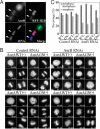
Aurora-AG198N prevents chromosome misalignment and cell premature exit from mitosis caused by Aurora-B knockdown. (A) Immunofluorescence photomicrographs of HeLa cells cotransfected with Aurora-B RNAi and RFP-H2B (red) at a ratio of 40:1 and labeled with Aurora-B antibody (green). Note that those cells expressing RFP-H2B showed disperse staining of Aurora-B with decreased fluorescence intensity, as indicated by arrows. Thus, in addition to chromosome morphology, RFP-H2B was a marker for Aurora-B knockdown. DNA labeled with DAPI (blue). (B) HeLa cells induced by tetracycline to express Aurora-AWT or Aurora-AG198N were cotransfected with Aurora-B RNAi and RFP-H2B at a ratio of 40:1, and subjected to time-course fluorescence microscopy. Chromosome morphology, visualized by RFP-H2B staining, was filmed every 5 min. With Aurora-B knockdown, cells could not properly align the chromosomes and exited mitosis without chromosome separation and cytokinesis. Induced expression of Aurora-AG198N, but not Aurora-AWT, prevented this abnormality. Representative cells can be viewed as movies or still images in Movie S1, Movie S2, Movie S3, Movie S4, Movie S5, Movie S6, Movie S7, and Movie S8. (C) Quantitative characterization of mitotic HeLa cells treated as in B. Dark-gray bars represent the percentage of cells from prophase to metaphase, and light-gray bars represent the percentage in anaphase. Cells were classified by chromosome morphology visualized by RFP-H2B. Each data point represents 3 independent experiments, each measuring 250 mitotic cells, and error bars indicate SD (P < 0.01).
Fig. 3.
Fig. 3.
Aurora-AG198N physically interacts with INCENP and Survivin, and its localization depends on them. (A) GFP-tagged Aurora-AWT, Aurora-AG198N, or Aurora-BWT was transfected into HeLa cells and immunoprecipitated from cell lysate with GFP antibody. The immunoprecipitated complexes were blotted for INCENP, endogenous Aurora-B, Survivin, or GFP. Aurora-AG198N bound INCENP and Survivin, not Aurora-B. (B and C) Photomicrographs of tet-on HeLa cells transfected with INCENP RNAi (B) or Survivin RNAi (C) in the presence of tetracycline and labeled with INCENP (B) or Survivin (C) antibody. Knockdown of INCENP or Survivin displaced Aurora-AG198N from centromere and midzone. Arrows indicate lagging chromosomes. (Scale bars, 10 μm.) (D) Quantitative characterization of Aurora-AG198N localization after INCENP or Survivin RNAi. Dark-gray bars represent the percentage of cells positively stained with Aurora-AG198N at centromere and midzone, and light-gray bars represent the percentage unstained at these locations. Each data point represents 3 independent experiments, each measuring 250 mitotic cells, and error bars indicate SD (P < 0.01). (E) GST pull-down assay for recombinant Aurora-AWT, Aurora-AG198N, or Aurora-BWT using GST-INCENP826–919, in the presence or absence of GFP-TPX21–43. Aurora-AG198N bound INCENP826–919 regardless of TPX21–43 addition (Top and Middle). Although Aurora-AWT bound INCENP826–919 (Top), this interaction was remarkably reduced when TPX21–43 was added (Middle). Input proteins were shown in separate lane with corresponding amount (Bottom). Arrowheads indicate Aurora-AWT, Aurora-AG198N, and Aurora-BWT.
Fig. 4.
Fig. 4.
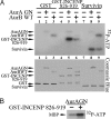
Aurora-AG198N phosphorylates INCENP and Survivin in vitro, and its activity is enhanced by INCENP. (A) In vitro kinase assay for recombinant Aurora-AG198N using GST, GST-INCENP826–919, and Survivin as substrates. Phosphorylated INCENP and Survivin were detected by autoradiography (Top), and total inputs were visualized by Coomassie blue staining (Middle). Lane 1–3, GST; lane 4–6, GST-INCENP826–919; lane 7–9, Survivin. Lanes 1, 4, and 7, without kinase; lanes 2, 5, and 8, with Aurora-AG198N; lanes 3, 6, and 9, with Aurora-BWT. (B) In vitro kinase assay for recombinant Aurora-AG198N using MBP as substrate, in the presence or absence of GST-INCENP826–919. Phosphorylated MBP was detected by autoradiography. INCENP826–919 stimulated phosphorylation of MBP by Aurora-AG198N.
Fig. 5.
Fig. 5.
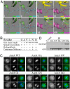
Both elongation and hydrophilicity of the side chain at residue 198 are required to convert Aurora-A into Aurora-B-like kinase. (A) Aurora-A and -B are distinguished by different partner proteins through local structural change at a single amino acid. (i and ii) The complex of human Aurora-A122–403/TPX21–43 (PDB ID code 1OL5 in ref. 14). (v and vi) The complex of Xenopus Aurora-B78–361/INCENP798–840 (PDB ID code 2BFY in ref. 37). (iii, iv, vii, and viii) Predicted by point mutation using PyMol (www.pymol.org). Structurally, G198 of Aurora-A (green) interacts with TPX2P13 (blue) in 3-dimensional space (i and ii). When G198 is replaced by N, the side chain is elongated, and TPX2 is prevented from Aurora-A (iii and iv). In contrast, Aurora-B (yellow) interacts with INCENP (purple) in a looser way within the equivalent domain (v and vi). The side chain of N158 in Xenopus Aurora-B interacts perfectly with INCENPY825/I828/D829/S830 and forms hydrogen bonds with INCENPY825/I828. Thus, Aurora-AG198N can interact with INCENP through its N198, which not only fits the 3-dimensional structure but probably builds hydrogen bonds with human INCENPI872 (vii and viii). (B) Overview of Aurora-A localization after substituting G198 with various amino acids. When the length of side chain reached that of V and L, Aurora-A was prevented from spindle. Hydrophilic side chains N and Q localized Aurora-A to centromere and midzone. (C) HeLa cells were transfected with GFP-tagged Aurora-AWT, Aurora-AG198A, Aurora-AG198V, Aurora-AG198L, Aurora-AG198N, or Aurora-AG198Q (Left, and green in Right), and subjected to immunofluorescence microscopy. DNA labeled with DAPI (Center, and blue in Right). Arrows indicate centromere and midzone. (Scale bar, 10 μm.) (D) GFP-tagged Aurora-AG198A, Aurora-AG198V, Aurora-AG198L, Aurora-AG198N, or Aurora-AG198Q was transfected into HeLa cells and immunoprecipitated from cell lysate with GFP antibody. The immunoprecipitated complexes were blotted for INCENP (Upper) or GFP (Lower). Aurora-AG198N and Aurora-AG198Q bound INCENP, whereas Aurora-AG198A, Aurora-AG198V, and Aurora-AG198L did not.
Fig. 6.
Fig. 6.
Model depicting the relationship between Aurora-A and -B: Aurora-A substitutes for Aurora-B in mitosis when it is displaced to the location of Aurora-B. (A) Under conventional condition, Aurora-A (green) binds TPX21–43 (blue) and localizes to spindle microtubule, and Aurora-B (yellow) binds INCENP826–919 (purple) and localizes to centromere and midzone. (B) When Aurora-A is altered at G198 by N, it binds INCENP instead of TPX2. In this complex containing at least Aurora-AG198N, INCENP, and Survivin, Aurora-AG198N is activated and phosphorylates Aurora-B substrates. Hence, when Aurora-B is absent from the cell, Aurora-AG198N maintains normal progression of the cell cycle.

References

    1. Taylor S, Peters JM. Polo and Aurora kinases: Lessons derived from chemical biology. Curr Opin Cell Biol. 2008;20:77–84. - PubMed
    1. Fu J, Bian M, Jiang Q, Zhang C. Roles of Aurora kinases in mitosis and tumorigenesis. Mol Cancer Res. 2007;5:1–10. - PubMed
    1. Giet R, Petretti C, Prigent C. Aurora kinases, aneuploidy and cancer, a coincidence or a real link? Trends Cell Biol. 2005;15:241–250. - PubMed
    1. Meraldi P, Honda R, Nigg EA. Aurora kinases link chromosome segregation and cell division to cancer susceptibility. Curr Opin Genet Dev. 2004;14:29–36. - PubMed
    1. Keen N, Taylor S. Aurora-kinase inhibitors as anticancer agents. Nat Rev Cancer. 2004;4:927–936. - PubMed

Publication types

MeSH terms