. 2009 Jun;101(6):3192-8.
doi: 10.1152/jn.91111.2008. Epub 2009 Apr 8.

Stress-induced changes in nucleus accumbens glutamate synaptic plasticity

Affiliations

Stress-induced changes in nucleus accumbens glutamate synaptic plasticity

Matthew R Campioni et al. J Neurophysiol. 2009 Jun.

Abstract

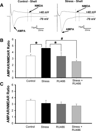
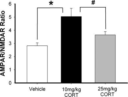
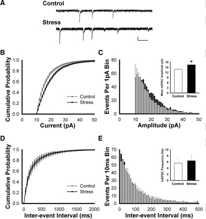
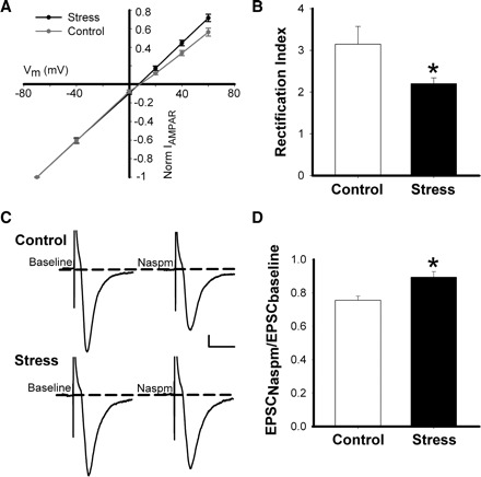
Stress hormones released in the CNS following exposure to unavoidable, aversive stimuli have been shown to alter the physiology of neurons in multiple brain regions including hippocampus, amygdala, prefrontal cortex, and ventral tegmental area. The nucleus accumbens (NAc), a motor-limbic interface linked to motivation and reward, receives inputs from each of these stress-affected brain regions, raising the possibility that its function might also be altered in response to stress. To assess potential stress-induced plasticity in the NAc, we exposed adult mice to daily cold water forced swim for 2 consecutive days and conducted electrophysiological experiments assessing glutamate receptor function in brain slices taken 18-24 h following the second swim. We found that AMPA receptor (AMPAR)/N-methyl-d-aspartate receptor (NMDAR) ratios, a measure of synaptic strength, were increased in the NAc shell but not core medium spiny neurons (MSNs) in stressed animals relative to controls. This effect was blocked by preadministration of glucocorticoid receptor (GR) antagonist RU486, suggesting that the observed changes are dependent on corticosteroid signaling. The role of corticosterone (CORT) in the observed plasticity was confirmed, because exogenous administration of 10 mg/kg CORT also enhanced AMPAR/NMDAR ratios in the NAc shell. The synaptic changes in NAc shell MSNs reflect an enhancement of AMPAR-mediated currents, as we observed increased AMPAR miniature postsynaptic current (mEPSC) amplitude following stress but no change in NMDAR mEPSCs. We hypothesize that altered information processing via plasticity of excitatory inputs might contribute to reward-related behaviors such as stress-induced reinstatement of drug seeking in animals and relapse in humans.


Figures

FIG. 1.
FIG. 1.
Stress increases AMPAR/NMDAR ratio in the nucleus accumbens (NAc) shell but not core. A: sample traces in control and stressed animals, showing how AMPAR/NMDAR ratios were obtained. B: averaged AMPAR/NMDAR ratios measured in the NAc shell from control animals, stressed animals, animals administered RU486, and animals administered RU486 30 min before cold water stress. The AMPAR/NMDAR ratios were increased in stressed animals compared with all other groups (*P < 0.02, #P < 0.0005). C: averaged AMPAR/NMDAR ratios measured in the NAc core under the same conditions as in B.
FIG. 2.
FIG. 2.
Corticosterone (CORT) increases AMPAR/NMDAR ratio of glutamatergic inputs to NAc shell medium spiny neurons (MSNs). Average AMPAR/NMDAR ratios in NAc shell MSNs measured in mice given 2 consecutive daily intraperitoneal injections of vehicle (50% DMSO, 50% saline), 10 mg/kg CORT, or 25 mg/kg CORT. The AMPAR/NMDAR ratios from animals receiving the 10 mg/kg CORT were increased compared with control and 25 mg/kg CORT-treated mice (*P < 0.003, #P < 0.035).
FIG. 3.
FIG. 3.
AMPAR miniature excitatory postsynaptic currents (mEPSCs) are larger in the NAc shell from stressed animals. A: sample traces showing typical mEPSCs at −70 mV. Scale bar: 10 pA, 100 ms. B: cumulative amplitude distributions of mEPSCs (1-pA bins) measured in the NAc shell MSNs, showing a shift toward larger events following stress relative to control (Kolmogorov-Smirnov test; P < 0.022). C: frequency histogram showing distribution of mEPSC amplitudes (1-pA bins) in control (crosshatched bars) and stressed (black bars) animals. Inset: average mEPSC amplitude is increased in the NAc shell of stressed animals relative to controls (t-test; P < 0.02). D: cumulative interevent interval distributions of mEPSCs (50-ms bins) in the NAc shell of control and stressed mice. E: frequency histogram showing distribution of mEPSC interevent intervals (10-ms bins). Inset: average mEPSC frequency is similar in NAc shell MSNs in control and stressed mice (t-test; P > 0.73).
FIG. 4.
FIG. 4.
Stress increases the linearity of the AMPAR receptor I-V relationship in NAc shell MSNs. A: AMPAR current-voltage relationship for neurons shown in B. B: rectification index (I70mV/I+40mV) showing reduced AMPAR current rectification in stressed animals relative to controls (*P < 0.05). C: sample traces showing the effects of 1-naphthylacetyl spermine trihydrochloride (Naspm; 100–200 μM) on evoked AMPAR EPSCs recorded at −70 mV from NAc MSNs of control and stressed mice. Scale bar: 25 pA, 30 ms. D: the effects of Naspm application on evoked AMPAR EPSCs reported as EPSC amplitude following 8 to 10 min drug wash in normalized to baseline EPSC amplitude. Naspm-mediated reduction of AMPAR EPSC amplitudes was greater in controls than in stressed mice (*P < 0.01).
FIG. 5.
FIG. 5.
Stress has no effect on NMDAR mEPSCs. A: sample traces of mEPSCs from NAc shell MSNs, recorded in 0 Mg2+ external solution, before and after d-AP5 (50 μM). Scale bar: 10 pA, 100 ms. B: example of averaged mEPSCs before (black) and after (dark gray) d-AP5 in a representative MSN, with the subtracted average NMDAR mEPSC (light gray). C: average of subtracted average NMDAR mEPSC amplitudes showing no difference between control and stressed mice (P < 0.5).

References

    1. Barrot M, Marinelli M, Abrous DN, Rouge-Pont F, Le Moal M, Piazza PV. Functional heterogeneity in dopamine release and in the expression of Fos-like proteins within the rat striatal complex. Eur J Neurosci 11: 1155–1166, 1999 - PubMed
    1. Barrot M, Marinelli M, Abrous DN, Rouge-Pont F, Le Moal M, Piazza PV. The dopaminergic hyper-responsiveness of the shell of the nucleus accumbens is hormone-dependent. Eur J Neurosci 12: 973–979, 2000 - PubMed
    1. Boudreau AC, Reimers JM, Milovanovic M, Wolf ME. Cell surface AMPA receptors in the rat nucleus accumbens increase during cocaine withdrawal but internalize after cocaine challenge in association with altered activation of mitogen-activated protein kinases. J Neurosci 27: 10621–10635, 2007 - PMC - PubMed
    1. Boudreau AC, Wolf ME. Behavioral sensitization to cocaine is associated with increased AMPA receptor surface expression in the nucleus accumbens. J Neurosci 25: 9144–9151, 2005 - PMC - PubMed
    1. Burnashev N, Monyer H, Seeburg PH, Sakmann B. Divalent ion permeability of AMPA receptor channels is dominated by the edited form of a single subunit. Neuron 8: 189–198, 1992 - PubMed

Publication types

MeSH terms

LinkOut - more resources