

Relationship between RPE and choriocapillaris in age-related macular degeneration
- PMID: 19357355
- PMCID: PMC4829357
- DOI: 10.1167/iovs.09-3639
Relationship between RPE and choriocapillaris in age-related macular degeneration
Abstract
Purpose: The purpose of this study was to examine the relationships between choriocapillaris (CC) and retinal pigment epithelial changes in age-related macular degeneration (AMD). Morphologic changes in the retinal pigment epithelium (RPE)/choriocapillaris complex were quantified in dry and wet forms of AMD, and the results were compared with those in aged control eyes without maculopathy.
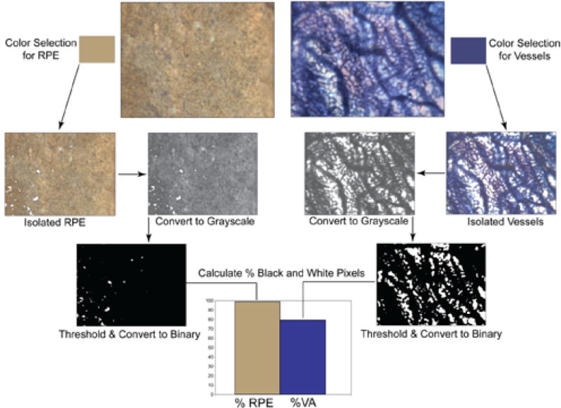
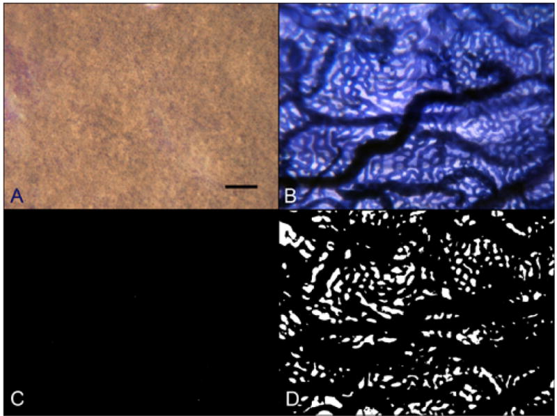
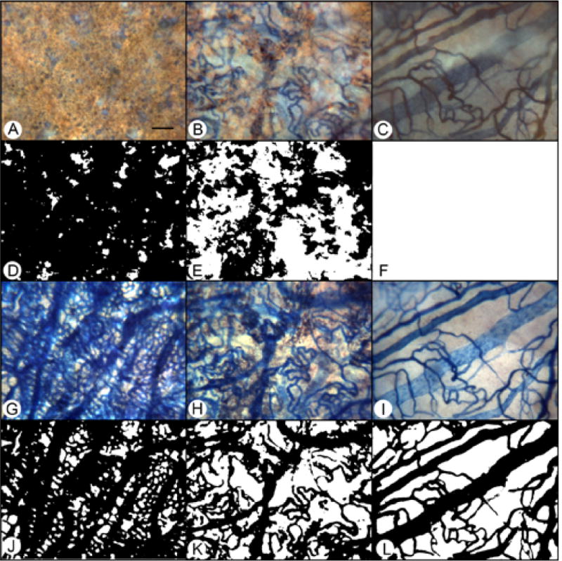
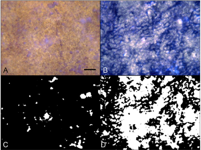
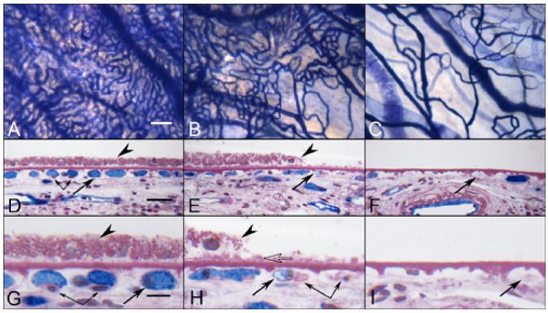
Methods: Postmortem choroids from three aged control subjects, five subjects with geographic atrophy (GA), and three subjects with wet AMD were analyzed using a semiquantitative computer-assisted morphometric technique developed to measure the percentages of retinal pigment epithelial and CC areas in choroidal wholemounts incubated for alkaline phosphatase activity. The tissues were subsequently embedded in methacrylate and were sectioned so that structural changes could be examined.
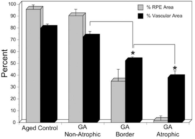
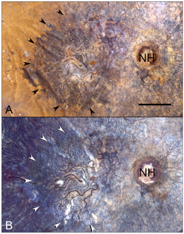
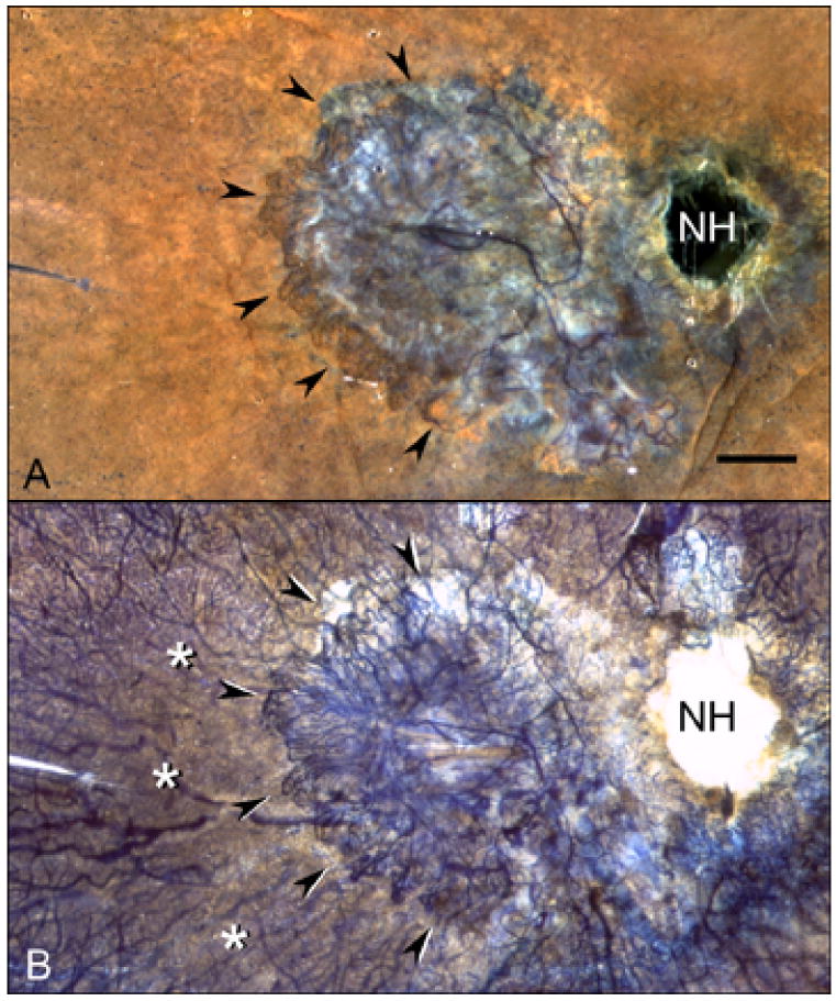
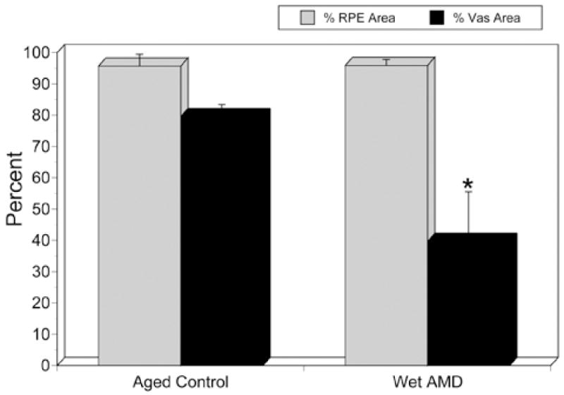
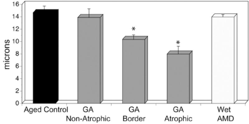
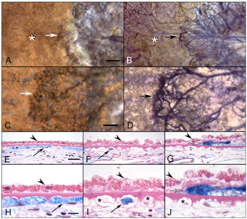
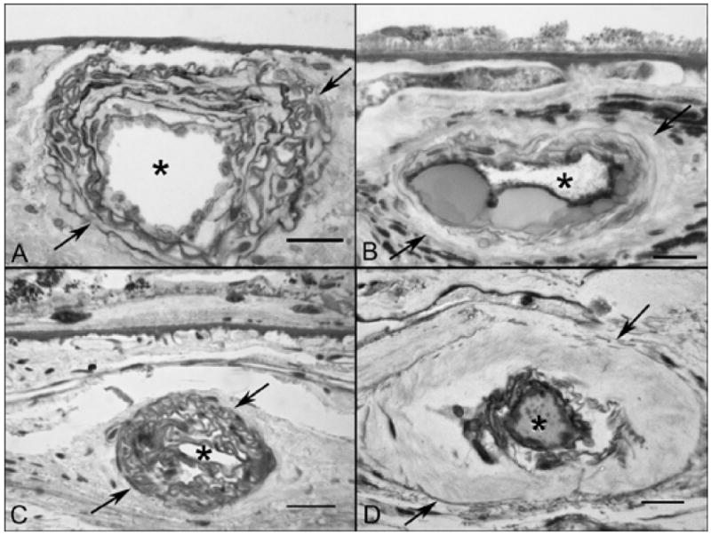
Results: There was a linear relationship between the loss of RPE and CC in GA. A 50% reduction in vascular area was found in regions of complete retinal pigment epithelial atrophy. Extreme constriction of remaining viable capillaries was found in areas devoid of RPE. Adjacent to active choroidal neovascularization (CNV) in wet AMD, CC dropout was evident in the absence of retinal pigment epithelial atrophy, resulting in a 50% decrease in vascular area. Lumenal diameters of the remaining capillaries in wet AMD eyes were similar to those in control eyes.
Conclusions: The primary insult in GA appears to be at the level of the RPE, and there is an intimate relationship between retinal pigment epithelial atrophy and secondary CC degeneration. CC degeneration occurs in the presence of viable RPE in wet AMD. The RPE in regions of vascular dropout are presumably hypoxic, which may result in an increase in VEGF production by the RPE and stimulation of CNV.
Figures
References
-
- Hayashi A, Majji AB, Fujioka S, Kim HC, Fukushima I, de Juan E., Jr Surgically induced degeneration and regeneration of the choriocapillaris in rabbit. Graefes Arch Clin Exp Ophthalmol. 1999;237:668–677. - PubMed
-
- Henkind P, Gartner S. The relationship between retinal pigment epithelium and the choriocapillaris. Trans Ophthalmol Soc U K. 1983;103:444–447. - PubMed
-
- Heriot WJ, Machemer R. Pigment epithelial repair. Graefes Arch Clin Exp Ophthalmol. 1992;230:91–100. - PubMed
-
- Korte GE, Repucci V, Henkind P. RPE destruction causes choriocapillary atrophy. Invest Ophthalmol Vis Sci. 1984;25:1135–1145. - PubMed
-
- Leonard DS, Sugino IK, Zhang XG, et al. Ultrastructural analysis of hydraulic and abrasive retinal pigment epithelial cell debridements. Exp Eye Res. 2003;76:473–491. - PubMed
Publication types
MeSH terms
Substances
Grants and funding
LinkOut - more resources
Full Text Sources
Other Literature Sources
Medical
