

Neuropathic pain in temporomandibular joint disorders: case-control analysis by MR imaging
- PMID: 19357384
- PMCID: PMC7051545
- DOI: 10.3174/ajnr.A1575
Neuropathic pain in temporomandibular joint disorders: case-control analysis by MR imaging
Abstract
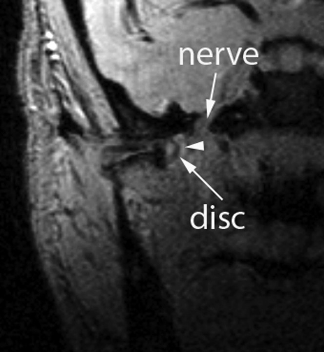
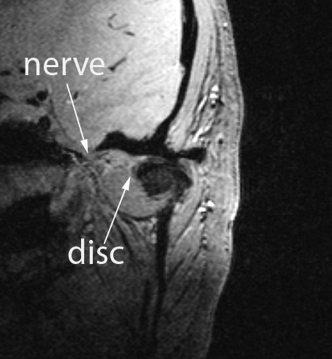
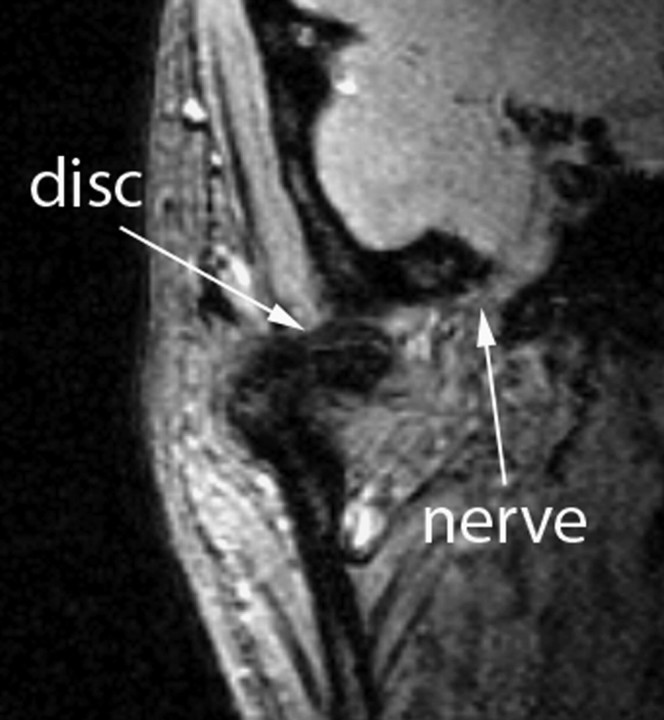
Background and purpose: Temporomandibular joint disorders (TMJ-D) may be associated with the onset of neuropathic pain. The purpose of this study was to prospectively assess if, at the open-mouth position, the distance between the temporomandibular joint (TMJ) disk and the mandibular nerve is shorter in patients with TMJ-D and neuropathic pain vs patients with TMJ-D without neuropathic pain or in healthy people.
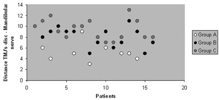
Materials and methods: After ethical committee approval, we evaluated by MR imaging 16 TMJs with TMJ-D and neuropathic pain, 16 TMJs with TMJ-D without neuropathic pain, and 16 TMJs of healthy volunteers. All of the subjects were informed about the study procedure. We evaluated the distance between the TMJ disk and the mandibular nerve at the oval foramen level. Furthermore, the presence within the TMJs of internal derangement, osteoarthrosis, joint effusion, and bone marrow edema was evaluated.
Results: At the maximal open-mouth position, the distance between the TMJ disk and the mandibular nerve is shorter in patients with TMJ-D and neuropathic pain than in patients with TMJ-D without neuropathic pain or in healthy volunteers (P < .05). The imaging findings of TMJ internal derangement, effusion, osteoarthrosis, and bone marrow edema were present both in patients with TMJ-D without neuropathic pain and in patients with TMJ-D and neuropathic pain.
Conclusions: We suggest that a closer proximity between the TMJ disk and the mandibular nerve could be one of the causes of the onset of neuropathic pain in patients with TMJ-D and neuropathic pain.
Figures
Similar articles
-
Relative odds of temporomandibular joint pain as a function of magnetic resonance imaging findings of internal derangement, osteoarthrosis, effusion, and bone marrow edema.Oral Surg Oral Med Oral Pathol Oral Radiol Endod. 2003 Apr;95(4):437-45. doi: 10.1067/moe.2003.95. Oral Surg Oral Med Oral Pathol Oral Radiol Endod. 2003. PMID: 12686927
-
Bone marrow edema of the mandibular condyle related to internal derangement, osteoarthrosis, and joint effusion.J Oral Maxillofac Surg. 2003 Jan;61(1):35-40. doi: 10.1053/joms.2003.50006. J Oral Maxillofac Surg. 2003. PMID: 12524605
-
Magnetic resonance imaging findings of internal derangement and effusion in patients with unilateral temporomandibular joint pain.Oral Surg Oral Med Oral Pathol Oral Radiol Endod. 2001 Nov;92(5):566-71. doi: 10.1067/moe.2001.116817. Oral Surg Oral Med Oral Pathol Oral Radiol Endod. 2001. PMID: 11709695
-
Role of magnetic resonance imaging in the clinical diagnosis of the temporomandibular joint.Cells Tissues Organs. 2005;180(1):6-21. doi: 10.1159/000086194. Cells Tissues Organs. 2005. PMID: 16088129 Review.
-
Association of bone marrow edema with temporomandibular joint (TMJ) osteoarthritis and internal derangements.Cranio. 2017 Jan;35(1):4-9. doi: 10.1080/08869634.2016.1156282. Epub 2016 Apr 8. Cranio. 2017. PMID: 27077262 Review.
Cited by
-
TMJ inferior compartment arthroplasty procedure through a 25-year follow-up (functional arthroplasty).Ann Stomatol (Roma). 2017 Jan 10;7(3):60-64. doi: 10.11138/ads/2016.7.3.060. eCollection 2016 Jul-Sep. Ann Stomatol (Roma). 2017. PMID: 28149452 Free PMC article.
-
Unlocking the potential of capsaicin in oral health (Review).Biomed Rep. 2024 Aug 22;21(5):153. doi: 10.3892/br.2024.1841. eCollection 2024 Nov. Biomed Rep. 2024. PMID: 39247424 Free PMC article. Review.
-
Thermal Effects of High-Intensity Laser Therapy on the Temporomandibular Joint Area in Clinically Healthy Racehorses-A Pilot Study.Animals (Basel). 2025 May 15;15(10):1426. doi: 10.3390/ani15101426. Animals (Basel). 2025. PMID: 40427303 Free PMC article.
-
Knee orthopedics as a template for the temporomandibular joint.Cell Rep Med. 2021 Apr 14;2(5):100241. doi: 10.1016/j.xcrm.2021.100241. eCollection 2021 May 18. Cell Rep Med. 2021. PMID: 34095872 Free PMC article. Review.
-
Complications of Mandibular Distraction Osteogenesis in Infants with Isolated Robin Sequence.Children (Basel). 2023 Sep 23;10(10):1591. doi: 10.3390/children10101591. Children (Basel). 2023. PMID: 37892254 Free PMC article.
References
-
- Okeson JP. Functional anatomy. In: Maino G. Management of Temporomandibular Disorders and Occlusion. 5th ed. Bologna, Italy: Edizioni Martina;2006. :1–127
-
- Dupont JS Jr. The prevalence of trigeminal neuritis with TMD. Cranio 2003;21:180–84 - PubMed
-
- Emshoff R, Brandlmaier I, Bertram S, et al. Relative odds of temporomandibular joint pain as a function of magnetic resonance imaging findings of internal derangement, osteoarthrosis, effusion, and bone marrow edema. Oral Surg Oral Med Oral Pathol Oral Radiol Endod 2003;95:437–45 - PubMed
-
- Ogura I. Magnetic resonance imaging characteristics of temporomandibular joint pain during opening and biting in patients with disc displacement. Oral Surg Oral Med Oral Pathol Oral Radiol Endod 2006;102:669–72 - PubMed
-
- Sano T, Westesson PL. Magnetic resonance imaging of the temporomandibular joint. Increased T2 signal in the retrodiscal tissue of painful joints. Oral Surg Oral Med Oral Pathol Oral Radiol Endod 1995;79:511–16 - PubMed
MeSH terms
LinkOut - more resources
Full Text Sources
Medical