. 2009 May;86(5):492-502.
doi: 10.1097/OPX.0b013e31819f9e4d.

Clinical and laboratory evaluation of peripheral prism glasses for hemianopia

Affiliations

Clinical and laboratory evaluation of peripheral prism glasses for hemianopia

Robert G Giorgi et al. Optom Vis Sci. 2009 May.

Abstract

Purpose: Homonymous hemianopia (the loss of vision on the same side in each eye) impairs the ability to navigate and walk safely. We evaluated peripheral prism glasses as a low vision optical device for hemianopia in an extended wearing trial.

Methods: Twenty-three patients with complete hemianopia (13 right) with neither visual neglect nor cognitive deficit enrolled in the 5-visit study. To expand the horizontal visual field, patients' spectacles were fitted with both upper and lower Press-On Fresnel prism segments (each 40 prism diopters) across the upper and lower portions of the lens on the hemianopic ("blind") side. Patients were asked to wear these spectacles as much as possible for the duration of the study, which averaged 9 (range: 5 to 13) weeks. Clinical success (continued wear, indicating perceived overall benefit), visual field expansion, perceived direction, and perceived quality of life were measured.

Results: Clinical success: 14 of 21 (67%) patients chose to continue to wear the peripheral prism glasses at the end of the study (two patients did not complete the study for non-vision reasons). At long-term follow-up (8 to 51 months), 5 of 12 (42%) patients reported still wearing the device. Visual field expansion: expansion of about 22 degrees in both the upper and lower quadrants was demonstrated for all patients (binocular perimetry, Goldmann V4e). Perceived direction: two patients demonstrated a transient adaptation to the change in visual direction produced by the peripheral prism glasses. Quality of life: at study end, reduced difficulty noticing obstacles on the hemianopic side was reported.

Conclusions: The peripheral prism glasses provided reported benefits (usually in obstacle avoidance) to 2/3 of the patients completing the study, a very good success rate for a vision rehabilitation device. Possible reasons for long-term discontinuation and limited adaptation of perceived direction are discussed.


Figures

Figure 1
Figure 1
Peripheral prism correction for a right hemianopic patient wearing spectacles with upper and lower 40Δ (22°) Press-On™ prism segments affixed base-out to the back of the right spectacle lens.
Figure 2
Figure 2
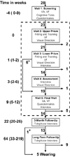
A flow diagram showing the timing of visits, the main procedures at each visit, and the numbers of patients. Numbers on the left of the flow diagram are the median time in weeks (range in parentheses) relative to the first fitting of a peripheral prism segment (upper at visit 2). Numbers next to the down arrows of the flow diagram show the numbers of patients moving to the next stage of the study. The numbers next to a right pointing arrow show the number of patients who discontinued wear of the peripheral prism glasses at each stage. Intentionally, six patients were not present at visit 4, shown by the asterisk. The “diamond” indicates the patient who had stopped wearing at 3-month follow-up, but reported wearing the peripheral prisms again at long-term follow-up. The formal visits of the study are enclosed by the dashed rectangle. VA = visual acuity; VF = Goldmann visual field.
Figure 3
Figure 3
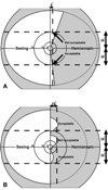
Illustration of the visual field inclusion criteria. The inclusion criteria were applied only to the area of the visual field between 30° above and below the horizontal midline. (a) Complete hemianopia test shown in a schematic binocular visual field. As the lower visual field extended less than 5° beyond the vertical midline within that zone, that quadrant was acceptable. However, the upper visual field extended more than 5° making this hypothetical patient ineligible for the study. (b) Homonymous hemianopia test illustrated with this schematic left eye monocular visual field. For this illustration, the right eye is assumed to have “ideal” hemianopia following the vertical meridian, hence a difference between the two eyes above or below 30° lines extending greater than 10 degrees beyond the vertical midline was acceptable, whereas the same difference within 30 degrees of the horizontal midline was not acceptable. A difference between the two eyes extending less than 10 degrees from the vertical midline in the latter zone was acceptable. The dotted lines in A and B represent normally-sighted binocular and monocular visual fields, respectively.
Figure 4
Figure 4
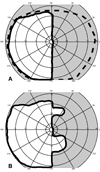
(a) Binocular visual field of a patient with right homonymous hemianopia is shown enclosed by the thick solid line. For comparison, the dashed line shows the binocular visual field of a normally-sighted subject. Stimuli presented in the grey portion were not seen by the patient. (b) Binocular visual field of the same patient wearing peripheral prism glasses. The difference in the visual field plots represents the visual field expansion provided by the peripheral prisms glasses. V4e target used for Goldmann kinetic perimetry.
Figure 5
Figure 5
Experimental set-up and apparatus for perceived direction testing. Patients sat 1 meter from a rear projection screen with their arms and hands occluded from view by use of a wooden box placed over a large bit pad on which patients could swing their arm, left and right. A brace was worn on the pointing arm to reduce flexion of the elbow. The patient’s task was to “point” to each seen stimulus and indicate its horizontal position by a mouse click. Stimulus locations are illustrated in Fig. 6. The background images were low contrast, grayscale images derived from cable television. For illustrative purposes, the contrast of this cartoon image here is higher and the size of the fixation target and circular stimulus greater than during actual perceived direction testing.
Figure 6
Figure 6
Perceived direction testing: Patients were presented twelve stimuli in each of eight zones, four zones in the hemianopic side (here left of vertical midline, 2 upper and 2 lower) and four zones in the seeing side (here right of vertical midline, 2 upper and 2 lower). Stimuli presented in zones “A” and “D” were detected in the seeing hemifield and were expected to be reported by the patient in the real direction. Stimuli presented in zones “B” were not expected to be detected by the patient, as these were presented in the hemianopic hemifield not covered by prism. Stimuli presented in zones “C” were expected to be detected through the prism segments shown as dashed lines and reported in the visual direction, demonstrating no adaptation of perceived direction, or in the real direction, demonstrating adaptation of perceived direction. The “X” represents the fixation target.
Figure 7
Figure 7
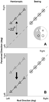
Idealized schematic data showing “no adaptation” and “adaptation” of perceived direction with prism segments worn by a patient with left hemianopia. (a) Stimuli detected in the seeing hemifield are reported in the real direction (i.e., the patient points correctly to the stimulus presented on the screen). Stimuli presented in the hemianopic hemifield and detected through prism segments have a visual direction that is shifted about 22° relative to the real direction by the 40Δ prisms. The perceived direction is the visual direction indicating no adaptation to the prism shift. (b) Stimuli seen in the seeing hemifield are reported as in (a), but stimuli seen through prism segments are reported in the real direction despite the prism shift of visual direction, showing adaptation of perceived direction.
Figure 8
Figure 8
Perceived direction of stimuli presented to the upper half of both the seeing and hemianopic hemifields for one patient with left hemianopia. (a) Results on the day of the upper prism segment fitting show no adaptation to the prism-induced change in visual direction. Stimuli seen through the prism segment (triangles, C Upper in fig. 6) were perceived about 22° to the right of the real direction, the expected deviation for the 40Δ power of the segment. Three of these stimuli, presented close to the hemifield border were detected by “peeking” into the hemianopic hemifield (dashed ellipse). Stimuli presented to the hemianopic hemifield in the non-prism zone (diamonds, B Upper) were not seen except for one stimulus again detected by “peeking” (dashed ellipse). Stimuli presented to corresponding zones in the seeing hemifield (squares, D Upper and circles, A Upper) were perceived in the real direction. (b) Results eight days after the upper prism segment fitting in which the patient reported wearing for an average of six hours per day. For stimuli seen through the prism segment the patient reported the stimuli direction as close to the real direction, demonstrating an “adaptation-like” response. However, the patient reported that he was making conscious, compensatory motor responses, which in (c), another eight days later are no longer reliably demonstrated. No adaptation was seen at visit 5 (data not shown).
Figure A1
Figure A1
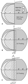
Expected binocular visual fields (white regions) of a person with right homonymous hemianopia with a 10° monocular sector prism worn over the right eye. In this figure it is assumed that the left eye maintains fixation and that the patient remains orthophoric. The direction of visual axis of the eye relative to the prism apex (altered by by turning the head left while maintaining fixation) is shown at the top of each panel. Binocular overlap occurs in the cross-hatched regions. A. When not looking through the prism there is no effect on the binocular visual field and thus no expansion. B. When the visual axis is directed slightly (that is less than the prism power, here 10°) into the prism (here 6°) there is visual field expansion represented by the sliver of right eye vision to the right of fixation. Note the narrow non-seeing gap (scotoma) between two seeing segments of the binocular visual field. There is also foveal double vision (visual confusion). C. When the visual axis is directed farther than the angle equivalent to the power of the prism towards the prism base (here 15°) there is visual field expansion with no gap. There is foveal double vision (both visual confusion and diplopia in this case).
Figure A2
Figure A2
Measured binocular visual fields of a patient with right hemianopia wearing a 9° monocular sector prism for the first time. A. Binocular visual field measured without the prism. The same field was measured when the patient was looking through the prism free part of the lenses. B. Binocular visual field measured with the patient looking slightly into the prism. This was achieved by tilting the head slightly to the left while maintaining fixation of the perimetry fixation target. A small visual field expansion to the right was recorded, together with a central optical scotoma (gap). C. With further shift of the gaze into the prism, visual field expansion with no gap was recorded. This visual field expansion is associated, however, with central double vision.
Figure A3
Figure A3
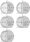
When, as reported, the “momentary” double vision disappears, the various ways it may be eliminated result in different visual field outcomes. Here, too, it is assumed that the patient remains orthophoric. A. If the right eye is suppressed completely, the binocular visual field remains about the same as that recorded without prism as the left eye visual field is not affected by the right lens prism. B. If the left eye is suppressed completely, the visual field is reduced to the nasal visual field of the right eye. Note the prism scotoma and the lack of expansion into the blind hemifield, since the right eye takes up fixation. C. If only central vision of the right eye is suppressed (again the left eye is assumed to be fixating), the visual field will be expanded, above and below fixation, into the blind hemifield. In this case as well as in D the peri-central areas of binocular overlap represent areas of double vision. D. If only central vision of the left eye is suppressed, since the right eye takes up fixation, the visual field will be expanded to the left, but not into the blind hemifield. E. If the user is able to fuse the double images seen through the 10° prism, the effect of the prism on the binocular visual field is eliminated. Note the lack of expansion into the blind hemifield, since both eyes fixate the target.

References

    1. Zhang X, Kedar S, Lynn MJ, Newman NJ, Biousse V. Homonymous hemianopia: Clinical-anatomic correlations in 904 cases. Neurology. 2006;66:906–910. - PubMed
    1. Williams GR, Jiang JG, Matchar DB, Samsa GP. Incidence and occurrence of total (first-ever and recurrent) stroke. Stroke. 1999;30:2523–2528. - PubMed
    1. Rossi PW, Kheyfets S, Reding J. Fresnel prisms improve perception in stroke patients with homonymous hemianopia or unilateral visual neglect. Neurology. 1990;40:1597–1599. - PubMed
    1. Zhang X, Kedar S, Lynn MJ, Newman NJ, Biousse V. Natural history of homonymous hemianopia. Neurology. 2006;66:901–905. - PubMed
    1. Kerkhoff G, Munssinger U, Haaf E, Eberlestrauss G, Stogerer E. Rehabilitation of homonymous scotomas in patients with postgeniculate damage of the visual-system-saccadic compensation training. Restor Neurol Neurosci. 1992;4:245–254. - PubMed

Publication types