. 2009;4(4):e5275.
doi: 10.1371/journal.pone.0005275. Epub 2008 Apr 16.

Pharmacologic inhibition of the TGF-beta type I receptor kinase has anabolic and anti-catabolic effects on bone

Affiliations

Pharmacologic inhibition of the TGF-beta type I receptor kinase has anabolic and anti-catabolic effects on bone

Khalid S Mohammad et al. PLoS One. 2009.

Abstract

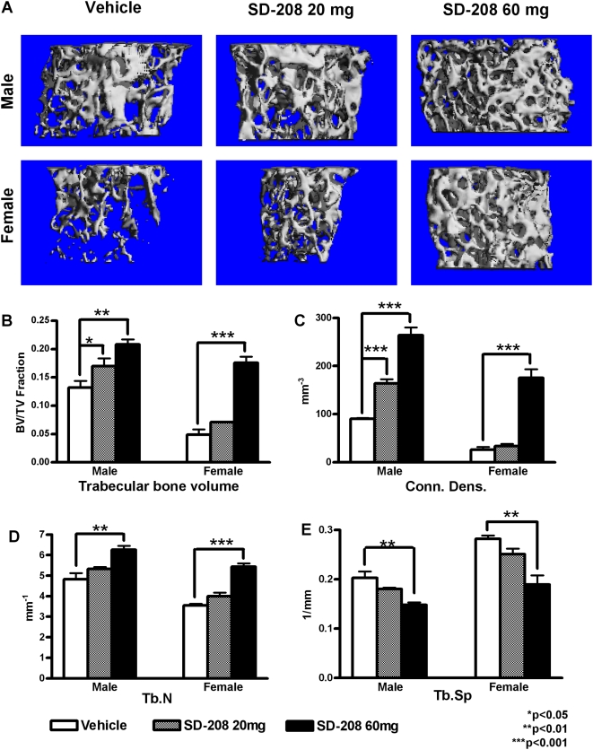
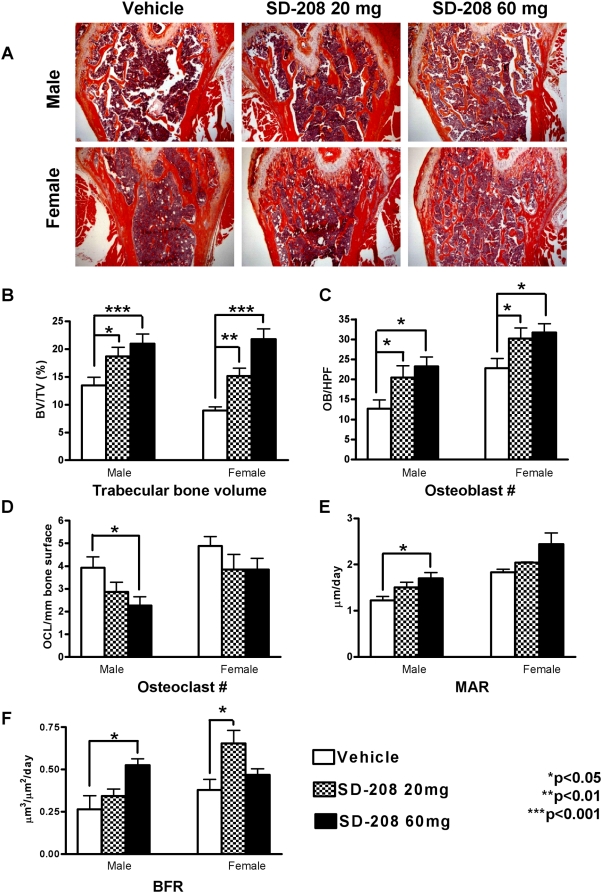
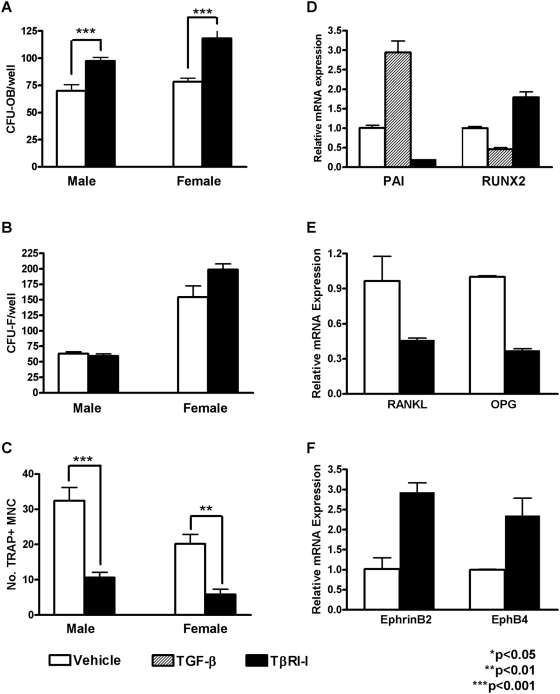
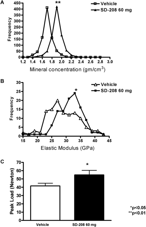
During development, growth factors and hormones cooperate to establish the unique sizes, shapes and material properties of individual bones. Among these, TGF-beta has been shown to developmentally regulate bone mass and bone matrix properties. However, the mechanisms that control postnatal skeletal integrity in a dynamic biological and mechanical environment are distinct from those that regulate bone development. In addition, despite advances in understanding the roles of TGF-beta signaling in osteoblasts and osteoclasts, the net effects of altered postnatal TGF-beta signaling on bone remain unclear. To examine the role of TGF-beta in the maintenance of the postnatal skeleton, we evaluated the effects of pharmacological inhibition of the TGF-beta type I receptor (TbetaRI) kinase on bone mass, architecture and material properties. Inhibition of TbetaRI function increased bone mass and multiple aspects of bone quality, including trabecular bone architecture and macro-mechanical behavior of vertebral bone. TbetaRI inhibitors achieved these effects by increasing osteoblast differentiation and bone formation, while reducing osteoclast differentiation and bone resorption. Furthermore, they induced the expression of Runx2 and EphB4, which promote osteoblast differentiation, and ephrinB2, which antagonizes osteoclast differentiation. Through these anabolic and anti-catabolic effects, TbetaRI inhibitors coordinate changes in multiple bone parameters, including bone mass, architecture, matrix mineral concentration and material properties, that collectively increase bone fracture resistance. Therefore, TbetaRI inhibitors may be effective in treating conditions of skeletal fragility.


Conflict of interest statement

Competing Interests: The authors have declared that no competing interests exist.

Figures

Figure 1
Figure 1. SD-208 inhibition of TGF-β function in vivo.
Five hours after TGF-β administration, SBE-Luc mice showed increased bioluminescence on the dorsal and ventral surfaces of the head where relatively little superficial tissue covers skeletal elements (calvarial bone and jaws) (a). Mice pretreated with SD-208 showed less basal and TGF-β-inducible luminescence than vehicle-treated controls (a, lower panels). SD-208 also inhibited reporter activity in SBE-Luc mouse calvarial explants cultured overnight with TGF-β (b). SD-208 treatment of calvarial explants inhibits expression of the TGF-β-inducible gene, PAI-1 , but induces expression of Runx2 and osteocalcin, osteoblast marker genes that are targets of TGF-β repression .
Figure 2
Figure 2. Pharmacologic TβRI inhibition increases BMD.
DXA was used to measure BMD longitudinally for male (a, c, e, g) and female mice (b, d, f, h) treated with or without the TβRI inhibitor SD-208 at 20 mg/kg or 60 mg/kg. SD-208 treatment at the 60 mg/kg dose caused an increase in total body (a, b) tibia (c, d), femur (e, f), and lumbar spine (g, h) BMD. SD-208 at the 20 mg/kg dose increased femoral BMD in female mice (f). Data represent mean±SEM (p<0.05, as determined by two-way analysis of variance (ANOVA).
Figure 3
Figure 3. Pharmacologic TβRI inhibition increases trabecular bone volume.
Micro-CT images show increased femoral trabecular bone volume following SD-208 treatment in male and female mice, relative to vehicle-treated controls (a). Quantitative analyses show that SD-208 increased trabecular bone volume (BV/TV, fraction) (b), connectivity density (c), and trabecular number (d), but decreased trabecular spacing (e) in male and female femora. Data represent mean±SEM (p<0.05, as determined by one-way ANOVA Newman-Keuls multiple comparison test).
Figure 4
Figure 4. Pharmacologic TβRI inhibition increases osteoblast numbers but reduces osteoclast numbers.
Representative H&E stained sections of femoral bone show the SD-208-dependent increase in trabecular bone in male and female mice (a). Histomorphometry shows that SD-208 increases trabecular bone volume in the femur (b) and tibia (data not shown), as well as osteoblast number (c) in a dose-dependent manner for male and female mice. Osteoclast numbers are reduced by SD-208 (60 mg/kg) in male mice (d). Dynamic histomorphometry of male mouse lumbar vertebrae shows that SD-208 treatment (60 mg/kg) increased mineral apposition rate (MAR) (e) and bone formation rate (BFR) (f). Data represent mean±SEM (*p<0.05, **p<0.01, ***p<0.001, as determined by one-way ANOVA Newman-Keuls multiple comparison test).
Figure 5
Figure 5. TβRI inhibition promotes osteoblast differentiation and bone deposition but inhibits osteoclast differentiation.
Bone marrow isolated from male and female mice treated with SD-208 (60 mg/kg) has increased numbers of osteoblast colony forming units (CFU-OB) (a) with no change in the number of colony forming units (CFU-F) (b). The number of TRAP-positive multinucleated cells (TRAP+ MNC) is lower in cultures from SD-208 treated mice than from vehicle-treated controls (c). Primary calvarial osteoblasts treated with TβRI-inhibitor SB431542 (10µM) or vehicle for 48 h show altered mRNA expression of PAI-1 (d) and several osteoblast and osteoclast regulatory factors including Runx2 (d), RANKL and OPG (e), and ephrinB2 and EphB4 (f). Data represent mean±SEM (*p<0.05, **p<0.01, ***p<0.001, as determined by unpaired t-test).
Figure 6
Figure 6. TβRI inhibitors increase bone mechanical and material properties.
Analysis of each pixel from XTM scans of femora show that SD-208 (60 mg/kg) increases bone matrix mineral concentration with a mean of 1.90 g/cm3+/−0.066, relative to a mean mineral concentration of 1.54 g/cm3+/−0.069 for vehicle-treated controls (p<0.05, as determined by unpaired t-test) (a). Analysis of elastic modulus values from nanoindents applied to tibial cortical bone showed a similar shift (p<0.05) (b). Unconfined compression testing of vertebrae from male mice treated with vehicle or SD-208 (60 mg/kg) shows an increased peak load-to-failure following TβRI inhibition (p<0.05) (e).

References

    1. Currey JD. The design of mineralised hard tissues for their mechanical functions. J Exp Biol. 1999;202:3285–3294. - PubMed
    1. Krishnan V, Bryant HU, Macdougald OA. Regulation of bone mass by Wnt signaling. J Clin Invest. 2006;116:1202–1209. - PMC - PubMed
    1. Wan M, Cao X. BMP signaling in skeletal development. Biochem Biophys Res Commun. 2005;328:651–657. - PubMed
    1. Alliston T, Piek E, Derynck R. TGF-beta family in skeletal development and disease. In: Derynck R, Miyazono K, editors. The TGF-β Family. Woodbury, NY: Cold Spring Harbor Press; 2008. pp. 667–723.
    1. Geiser AG, et al. Decreased bone mass and bone elasticity in mice lacking the transforming growth factor-beta1 gene. Bone. 1998;23:87–93. - PubMed

Publication types

MeSH terms

Substances